28
views
0
recommends
+1 Recommend
0 collections
    0
    shares
      • Record: found
      • Abstract: found
      • Article: not found

      Activation induced morphological changes and integrin αIIbβ3 activity of living platelets

      research-article

      Read this article at

      ScienceOpenPublisherPMC
      Bookmark
          There is no author summary for this article yet. Authors can add summaries to their articles on ScienceOpen to make them more accessible to a non-specialist audience.

          Highlights

          • We show stress induced acceleration of platelets shape changes.

          • Time-lapse AFM imaging visualizes two phases of filopodia extrusion and contraction.

          • SMFS reveals a close to 100 percent activity of the integrin αIIbβ3.

          Abstract

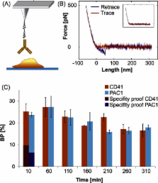
          Platelets are essential in hemostasis. Upon activation they undergo a shape-change accompanied with receptor presentation. Atomic force microscopy (AFM) imaging and single molecule force spectroscopy (SMFS) were used as powerful tools for exploring morphological changes as well as receptor activities of platelets. Imaging time series was accomplished with and without fixation steps at the single platelet level. Hereby the response of mechanical stimulation of the platelet by the AFM cantilever tip was directly observed. We demonstrate that living and fixed platelets develop filopodia after a short activation time followed by their disappearance including cellular bleb formation. Thereafter a second filopodia formation (filopodia extrusion) was observed; those filopodia subsequently disappeared again, and finally platelets detached from the support due to cell death. We determined the influence of mechanical stress on the chronology of morphological changes of platelets and demonstrated shear force induced filopodia formation. Through recordings over several hours, topographical AFM images over the full platelet lifetime – from early activation up to apoptosis – are presented. SMFS measurements on living platelets allowed determining the activation state of the most prominent membrane receptor integrin αIIbβ3 at all different phases of activation. αIIbβ3 was fully activated, independent of the morphological state.

          Related collections

          Most cited references39

          • Record: found
          • Abstract: found
          • Article: not found

          Platelets: physiology and biochemistry.

          Platelets are specialized blood cells that play central roles in physiologic and pathologic processes of hemostasis, inflammation, tumor metastasis, wound healing, and host defense. Activation of platelets is crucial for platelet function that includes a complex interplay of adhesion and signaling molecules. This article gives an overview of the activation processes involved in primary and secondary hemostasis, for example, platelet adhesion, platelet secretion, platelet aggregation, microvesicle formation, and clot retraction/stabilization. In addition, activated platelets are predominantly involved in cross talk to other blood and vascular cells. Stimulated "sticky" platelets enable recruitment of leukocytes at sites of vascular injury under high shear conditions. Platelet-derived microparticles as well as soluble adhesion molecules, sP-selectin and sCD40L, shed from the surface of activated platelets, are capable of activating, in turn, leukocytes and endothelial cells. This article focuses further on the new view of receptor-mediated thrombin generation of human platelets, necessary for the formation of a stable platelet-fibrin clot during secondary hemostasis. Finally, special emphasis is placed on important stimulatory and inhibitory signaling pathways that modulate platelet function.
            Bookmark
            • Record: found
            • Abstract: found
            • Article: not found

            Measuring the viscoelastic properties of human platelets with the atomic force microscope.

            We have measured force curves as a function of the lateral position on top of human platelets with the atomic force microscope. These force curves show the indentation of the cell as the tip loads the sample. By analyzing these force curves we were able to determine the elastic modulus of the platelet with a lateral resolution of approximately 100 nm. The elastic moduli were in a range of 1-50 kPa measured in the frequency range of 1-50 Hz. Loading forces could be controlled with a resolution of 80 pN and indentations of the platelet could be determined with a resolution of 20 nm.
              Bookmark
              • Record: found
              • Abstract: found
              • Article: not found

              Linking of Sensor Molecules with Amino Groups to Amino-Functionalized AFM Tips

              The measuring tip of an atomic force microscope (AFM) can be upgraded to a specific biosensor by attaching one or a few biomolecules to the apex of the tip. The biofunctionalized tip is then used to map cognate target molecules on a sample surface or to study biophysical parameters of interaction with the target molecules. The functionality of tip-bound sensor molecules is greatly enhanced if they are linked via a thin, flexible polymer chain. In a typical scheme of tip functionalization, reactive groups are first generated on the tip surface, a bifunctional cross-linker is then attached with one of its two reactive ends, and finally the probe molecule of interest is coupled to the free end of the cross-linker. Unfortunately, the most popular functional group generated on the tip surface is the amino group, while at the same time, the only useful coupling functions of many biomolecules (such as antibodies) are also NH2 groups. In the past, various tricks or detours were applied to minimize the undesired bivalent reaction of bifunctional linkers with adjacent NH2 groups on the tip surface. In the present study, an uncompromising solution to this problem was found with the help of a new cross-linker (“acetal-PEG-NHS”) which possesses one activated carboxyl group and one acetal-protected benzaldehyde function. The activated carboxyl ensures rapid unilateral attachment to the amino-functionalized tip, and only then is the terminal acetal group converted into the amino-reactive benzaldehyde function by mild treatment (1% citric acid, 1–10 min) which does not harm the AFM tip. As an exception, AFM tips with magnetic coating become demagnetized in 1% citric acid. This problem was solved by deprotecting the acetal group before coupling the PEG linker to the AFM tip. Bivalent binding of the corresponding linker (“aldehyde-PEG-NHS”) to adjacent NH2 groups on the tip was largely suppressed by high linker concentrations. In this way, magnetic AFM tips could be functionalized with an ethylene diamine derivative of ATP which showed specific interaction with mitochondrial uncoupling protein 1 (UCP1) that had been purified and reconstituted in a mica-supported planar lipid bilayer.
                Bookmark

                Author and article information

                Journal
                Methods
                Methods
                Methods (San Diego, Calif.)
                Academic Press
                1046-2023
                1095-9130
                01 April 2013
                01 April 2013
                : 60
                : 2
                : 179-185
                Affiliations
                Institute of Biophysics, University of Linz, Gruberstrasse 40, 4020 Linz, Austria
                Department of Internal Medicine II: Cardiology, Hospital of the “Barmherzige Schwestern”, Seilerstätte 4, 4010 Linz, Austria
                Department of Blood Group Serology and Transfusion Medicine, Medical University Vienna, Währinger Gürtel 18-20, 1090 Wien, Austria
                Author notes
                [* ]Corresponding author. andreas.ebner@ 123456jku.at
                Article
                YMETH3101
                10.1016/j.ymeth.2013.03.034
                3678024
                23571313
                725ae3bc-4e1e-4a82-baaa-8ca15ed93e26
                © 2013 The Authors

                This document may be redistributed and reused, subject to certain conditions.

                History
                Categories
                Article

                Life sciences
                platelets,single molecule force spectroscopy,atomic force microscopy,integrin αiibβ3

                Comments

                Comment on this article