4
views
0
recommends
+1 Recommend
0 collections
    0
    shares
      • Record: found
      • Abstract: found
      • Article: found
      Is Open Access

      Comparison of 99mTc radiolabeled somatostatin antagonist with [ 68 Ga]Ga-DOTA-TATE in a patient with advanced neuroendocrine tumor

      brief-report

      Read this article at

      Bookmark
          There is no author summary for this article yet. Authors can add summaries to their articles on ScienceOpen to make them more accessible to a non-specialist audience.

          Abstract

          Gallium-68-labelled somatostatin analogues are a gold standard of neuroendocrine tumors (NETs) PET imaging being a tool for personalized treatment. However, in some cases of low somatostatin receptor (SSTR) expressing tumors, its clinical value can be limited. SSTR antagonists in comparison to currently used agonistic analogues recognize more binding sites on NET cells [1], which may improve the diagnostic efficacy enabling more precise staging leading to the better outcome. SPECT radiopharmaceuticals represent the cornerstone of molecular imaging due to their wide availability [2] and the development of a radiopharmaceutical based on technetium-99 m-labeled SSTR antagonist would improve access to such clinically feasible diagnostic tool. 
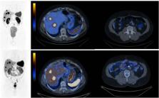
Below (Fig. 1), we present a comparison of SPECT/CT with a SSTR antagonist [99mTc]Tc-TECANT1 (N4-p-Cl-Phe-cyclo(D-Cys-Tyr-D-Aph(Cbm)-Lys-Thr-Cys)-D-Tyr-NH2, where D-Aph(Cbm): D-4-amino-carbamoyl-phenylalanine) [3] and [68 Ga]Ga-DOTA-TATE PET/CT (EudraCT no 2019–003379-20). Please note better visualization of the lesions seen in the study with [99mTc]Tc-TECANT1 (in 6 out of 7 lesions) as well as significantly higher tumor-to-background ratio (in primary and metastatic lesions) obtained with the novel tracer (measured as an absolute number of counts in lesion to background (TBR)) in comparison to [68 Ga]Ga-DOTA-TATE PET/CT (4,07 (range 1,36–7,58) vs 2,26 (range 1,15–3,39)). Fig. 1 Maximum intensity projection (MIP) and axial fused images: upper line -[99mTc]Tc-TECANT1 SPECT/CT (4 h post injection, injected activity 785 MBq, 120 frames, 20 s per frame); bottom line — [68 Ga]Ga-DOTA-TATE PET/CT (1 h post injection, injected activity 146 MBq; 3 min per bed). Scans were obtained within 13 days. Long-acting somatostatin analogue was withdrawn 4 weeks before imaging Although PET is considered more sensitive than SPECT [4], the presented new radiopharmaceutical holds promise to provide higher TBR values compared to the current gold standard 68 Ga-DOTA-TATE PET/CT. In combination with the development of quantitative SPECT imaging of NETs, the use of 99mTc-labelled SSTR antagonists could provide a widely available, clinically significant step in the personalized management of NETs.

          Related collections

          Most cited references4

          • Record: found
          • Abstract: found
          • Article: not found

          Somatostatin Receptor Antagonists for Imaging and Therapy.

          Somatostatin receptor (sstr) scintigraphy for imaging and sstr analogs for treatment have been used for more than 20 y. An important improvement in recent years was the introduction of peptide receptor radionuclide therapy with radiolabeled sstr agonists, such as [90Y-DOTA0,Tyr3]octreotide or [177Lu-DOTA0,Tyr3]octreotide (90Y- or177Lu-DOTATOC, respectively) and [177Lu-DOTA0,Tyr3]octreotate (177Lu-DOTATATE). PET/CT with68Ga-labeled sstr agonists, such as68Ga-DOTATOC,68Ga-DOTATATE, and [68Ga-DOTA,1-Nal3]octreotide (68Ga-DOTANOC), plays an important role in staging and restaging neuroendocrine tumors. Most importantly, sstr scintigraphy and sstr PET/CT can distinguish patients who will qualify for and benefit from peptide receptor radionuclide therapy. This characteristic of sstr targeting is important because it allows a personalized treatment approach (theranostic approach). Until recently, it was thought that internalization of the radiolabeled agonist was mandatory for sstr-mediated imaging and therapy. It was Ginj et al. who proposed in 2006 the paradigm shift that radiolabeled sstr antagonists may perform better than agonists despite the lack of internalization. Despite the rather limited number of head-to-head comparisons of sstr antagonists and agonists, the superiority of sstr antagonists was demonstrated in several cases. From a small library of sstr antagonists, the analog JR11 (Cpa-c[d-Cys-Aph(Hor)-d-Aph(Cbm)-Lys-Thr-Cys]-d-Tyr-NH2), an antagonist with selectivity for sstr subtype 2, showed the best overall characteristics for sstr subtype 2 targeting and was therefore selected for clinical translation. JR11 is under clinical development as a PET imaging agent when labeled with68Ga (68Ga-NODAGA-JR11 or68Ga-OPS202) and as a therapeutic agent when labeled with177Lu (177Lu-DOTA-JR11 or177Lu-OPS201). In this article, we discuss the development and current status of radiolabeled sstr antagonists. Evidence based on preclinical work, on quantitative in vivo autoradiography of human tumor slices, and on human data now supports a shift to sstr antagonists.
            Bookmark
            • Record: found
            • Abstract: not found
            • Book: not found

            The Supply of Medical Isotopes

            (2019)
              Bookmark
              • Record: found
              • Abstract: found
              • Article: not found

              Development of the 99mTc-Labelled SST2 Antagonist TECANT-1 for a First-in-Man Multicentre Clinical Study.

              Broad availability and cost-effectiveness of 99Mo/99mTc generators worldwide support the use, and thus the development, of novel 99mTc-labelled radiopharmaceuticals. In recent years, preclinical and clinical developments for neuroendocrine neoplasms patient management focused on somatostatin receptor subtype 2 (SST2) antagonists, mainly due to their superiority in SST2-tumour targeting and improved diagnostic sensitivity over agonists. The goal of this work was to provide a reliable method for facile preparation of a 99mTc-labelled SST2 antagonist, [99mTc]Tc-TECANT-1, in a hospital radiopharmacy setting, suitable for a multi-centre clinical trial. To ensure successful and reproducible on-site preparation of the radiopharmaceutical for human use shortly before administration, a freeze-dried three-vial kit was developed. The final composition of the kit was established based on the radiolabelling results obtained during the optimisation process, in which variables such as precursor content, pH and buffer, as well as kit formulations, were tested. Finally, the prepared GMP-grade batches met all predefined specification parameters together with long-term kit stability and stability of the product [99mTc]Tc-TECANT-1. Furthermore, the selected precursor content complies with micro-dosing, based on an extended single-dose toxicity study, where histopathology NOEL was established at 0.5 mg/kg BW, being more than 1000 times higher than the planned human dose of 20 µg. In conclusion, [99mTc]Tc-TECANT-1 is suitable to be advanced into a first-in-human clinical trial.
                Bookmark

                Author and article information

                Contributors
                alahub@cm-uj.krakow.pl
                Journal
                Eur J Nucl Med Mol Imaging
                Eur J Nucl Med Mol Imaging
                European Journal of Nuclear Medicine and Molecular Imaging
                Springer Berlin Heidelberg (Berlin/Heidelberg )
                1619-7070
                1619-7089
                15 July 2023
                15 July 2023
                2023
                : 50
                : 13
                : 4110-4111
                Affiliations
                [1 ]Chair and Department of Endocrinology, Jagiellonian University Medical College, ( https://ror.org/03bqmcz70) Krakow, Poland
                [2 ]Department of Nuclear Medicine, University Medical Centre Ljubljana, ( https://ror.org/01nr6fy72) Ljubljana, Slovenia
                [3 ]Faculty of Medicine, University of Ljubljana, ( https://ror.org/05njb9z20) Ljubljana, Slovenia
                [4 ]GRID grid.5361.1, ISNI 0000 0000 8853 2677, Department of Nuclear Medicine, , Medical University Innsbruck, ; Innsbruck, Austria
                [5 ]Faculty of Pharmacy, University of Ljubljana, ( https://ror.org/05njb9z20) Ljubljana, Slovenia
                [6 ]Radioisotope Centre POLATOM, National Centre for Nuclear Research, ( https://ror.org/00nzsxq20) Otwock, Poland
                [7 ]Faculty of Mathematics and Physics, University of Ljubljana, ( https://ror.org/05njb9z20) Ljubljana, Slovenia
                [8 ]Jožef Stefan Institute, ( https://ror.org/01hdkb925) Ljubljana, Slovenia
                [9 ]Universitätsspital Basel, ( https://ror.org/04k51q396) Basel, Switzerland
                [10 ]GRID grid.412700.0, ISNI 0000 0001 1216 0093, University Hospital Krakow, ; Krakow, Poland
                Author information
                http://orcid.org/0000-0001-5208-4689
                Article
                6335
                10.1007/s00259-023-06335-9
                10611881
                37452871
                73138717-e2eb-4da4-82af-bdc5fb0b0c42
                © The Author(s) 2023

                Open Access This article is licensed under a Creative Commons Attribution 4.0 International License, which permits use, sharing, adaptation, distribution and reproduction in any medium or format, as long as you give appropriate credit to the original author(s) and the source, provide a link to the Creative Commons licence, and indicate if changes were made. The images or other third party material in this article are included in the article's Creative Commons licence, unless indicated otherwise in a credit line to the material. If material is not included in the article's Creative Commons licence and your intended use is not permitted by statutory regulation or exceeds the permitted use, you will need to obtain permission directly from the copyright holder. To view a copy of this licence, visit http://creativecommons.org/licenses/by/4.0/.

                History
                : 2 June 2023
                : 6 July 2023
                Categories
                Image of the Month
                Custom metadata
                © Springer-Verlag GmbH Germany, part of Springer Nature 2023

                Radiology & Imaging
                Radiology & Imaging

                Comments

                Comment on this article