2
views
0
recommends
+1 Recommend
0 collections
    0
    shares
      • Record: found
      • Abstract: found
      • Article: found
      Is Open Access

      CAPTEM in Metastatic Well-Differentiated Intermediate to High Grade Neuroendocrine Tumors: A Single Centre Experience

      research-article

      Read this article at

      Bookmark
          There is no author summary for this article yet. Authors can add summaries to their articles on ScienceOpen to make them more accessible to a non-specialist audience.

          Abstract

          Introduction

          Capecitabine-temozolomide (CAPTEM) has significant activity in patients (pts) with metastatic low grade pancreatic neuroendocrine tumors (NETs). However, there is limited data regarding its activity in pts with metastatic well-differentiated intermediate and high grade pancreatic and nonpancreatic NETs. The objective of this study was to assess the functional imaging response, survival, and tolerability of CAPTEM in this population.

          Methods

          A retrospective audit of pts with metastatic well-differentiated intermediate (WHO grade 2) or high grade (WHO grade 3) NETs treated at Peter MacCallum Cancer Centre between March 2013 and March 2017. Pts received capecitabine 750 mg/m 2 orally twice daily (bd) from days1 to 14 and temozolomide 100 mg/m 2 bd from days 10 to 14 every 28 days. Data regarding functional imaging response, progression-free and overall survival, and toxicities was collected.

          Results

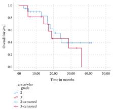
          Thirty-two pts received a median of 6 cycles (range: 2-16) of CAPTEM for grade 2 (n=21, 66%) or grade 3 (n=11, 34%), Ki67 <55% (n= 7, 21.9%) or Ki67 ≥55% (n= 4, 12.5 %) NET. Primary site included gastroenteropancreatic (n= 17, 53%), lung (n= 12, 37.5%), and unknown origin (n = 3, 9.4%). Twenty-two percent received CAPTEM as first-line therapy. After a median of 31 months of follow-up, the two-year overall survival (OS) was 42%, with a median OS of 24 months. There was a trend towards improved median progression-free survival (PFS) in pts with low grade 3 (Ki67<55%) versus high grade 3 (Ki67 ≥55%) NETs (15 vs 4 months, p= 0.11). Ten (31.3%) experienced grade 3/4 toxicity, with nausea (15.6%), thrombocytopaenia (12.5%), and fatigue (9.4%) the most common toxicities reported.

          Conclusion

          CAPTEM has significant activity in patients with metastatic grades 2 and 3 NETs with manageable toxicity. The PFS benefit observed in the grade 3 subgroup with Ki67<55% warrants further evaluation in a larger randomized trial.

          Related collections

          Most cited references34

          • Record: found
          • Abstract: found
          • Article: not found

          From RECIST to PERCIST: Evolving Considerations for PET response criteria in solid tumors.

          The purpose of this article is to review the status and limitations of anatomic tumor response metrics including the World Health Organization (WHO) criteria, the Response Evaluation Criteria in Solid Tumors (RECIST), and RECIST 1.1. This article also reviews qualitative and quantitative approaches to metabolic tumor response assessment with (18)F-FDG PET and proposes a draft framework for PET Response Criteria in Solid Tumors (PERCIST), version 1.0. PubMed searches, including searches for the terms RECIST, positron, WHO, FDG, cancer (including specific types), treatment response, region of interest, and derivative references, were performed. Abstracts and articles judged most relevant to the goals of this report were reviewed with emphasis on limitations and strengths of the anatomic and PET approaches to treatment response assessment. On the basis of these data and the authors' experience, draft criteria were formulated for PET tumor response to treatment. Approximately 3,000 potentially relevant references were screened. Anatomic imaging alone using standard WHO, RECIST, and RECIST 1.1 criteria is widely applied but still has limitations in response assessments. For example, despite effective treatment, changes in tumor size can be minimal in tumors such as lymphomas, sarcoma, hepatomas, mesothelioma, and gastrointestinal stromal tumor. CT tumor density, contrast enhancement, or MRI characteristics appear more informative than size but are not yet routinely applied. RECIST criteria may show progression of tumor more slowly than WHO criteria. RECIST 1.1 criteria (assessing a maximum of 5 tumor foci, vs. 10 in RECIST) result in a higher complete response rate than the original RECIST criteria, at least in lymph nodes. Variability appears greater in assessing progression than in assessing response. Qualitative and quantitative approaches to (18)F-FDG PET response assessment have been applied and require a consistent PET methodology to allow quantitative assessments. Statistically significant changes in tumor standardized uptake value (SUV) occur in careful test-retest studies of high-SUV tumors, with a change of 20% in SUV of a region 1 cm or larger in diameter; however, medically relevant beneficial changes are often associated with a 30% or greater decline. The more extensive the therapy, the greater the decline in SUV with most effective treatments. Important components of the proposed PERCIST criteria include assessing normal reference tissue values in a 3-cm-diameter region of interest in the liver, using a consistent PET protocol, using a fixed small region of interest about 1 cm(3) in volume (1.2-cm diameter) in the most active region of metabolically active tumors to minimize statistical variability, assessing tumor size, treating SUV lean measurements in the 1 (up to 5 optional) most metabolically active tumor focus as a continuous variable, requiring a 30% decline in SUV for "response," and deferring to RECIST 1.1 in cases that do not have (18)F-FDG avidity or are technically unsuitable. Criteria to define progression of tumor-absent new lesions are uncertain but are proposed. Anatomic imaging alone using standard WHO, RECIST, and RECIST 1.1 criteria have limitations, particularly in assessing the activity of newer cancer therapies that stabilize disease, whereas (18)F-FDG PET appears particularly valuable in such cases. The proposed PERCIST 1.0 criteria should serve as a starting point for use in clinical trials and in structured quantitative clinical reporting. Undoubtedly, subsequent revisions and enhancements will be required as validation studies are undertaken in varying diseases and treatments.
            Bookmark
            • Record: found
            • Abstract: found
            • Article: not found

            Predictive and prognostic factors for treatment and survival in 305 patients with advanced gastrointestinal neuroendocrine carcinoma (WHO G3): the NORDIC NEC study.

            As studies on gastrointestinal neuroendocrine carcinoma (WHO G3) (GI-NEC) are limited, we reviewed clinical data to identify predictive and prognostic markers for advanced GI-NEC patients. Data from advanced GI-NEC patients diagnosed 2000-2009 were retrospectively registered at 12 Nordic hospitals. The median survival was 11 months in 252 patients given palliative chemotherapy and 1 month in 53 patients receiving best supportive care (BSC) only. The response rate to first-line chemotherapy was 31% and 33% had stable disease. Ki-67<55% was by receiver operating characteristic analysis the best cut-off value concerning correlation to the response rate. Patients with Ki-67<55% had a lower response rate (15% versus 42%, P<0.001), but better survival than patients with Ki-67≥55% (14 versus 10 months, P<0.001). Platinum schedule did not affect the response rate or survival. The most important negative prognostic factors for survival were poor performance status (PS), primary colorectal tumors and elevated platelets or lactate dehydrogenase (LDH) levels. Advanced GI-NEC patients should be considered for chemotherapy treatment without delay.PS, colorectal primary and elevated platelets and LDH levels were prognostic factors for survival. Patients with Ki-67<55% were less responsive to platinum-based chemotherapy, but had a longer survival. Our data indicate that it may not be correct to consider all GI-NEC as one single disease entity.
              Bookmark
              • Record: found
              • Abstract: found
              • Article: found

              ENETS Consensus Guidelines for High-Grade Gastroenteropancreatic Neuroendocrine Tumors and Neuroendocrine Carcinomas

                Bookmark

                Author and article information

                Contributors
                Journal
                J Oncol
                J Oncol
                JO
                Journal of Oncology
                Hindawi
                1687-8450
                1687-8469
                2019
                20 February 2019
                : 2019
                : 9032753
                Affiliations
                1Department of Cancer Medicine, Peter MacCallum Cancer Centre, 305 Grattan Street, Melbourne, VIC 3000, Australia
                2Department Of Medical Oncology, Goulburn Valley Health, Shepparton VIC 3630, Australia
                3Department of Rural Health, Melbourne Medical School, The University of Melbourne, Shepparton, VIC, Australia
                4Neuroendocrine Unit, Peter MacCallum Cancer Centre, 305 Grattan St, Melbourne VIC 3000, Australia
                5Department of Cancer Imaging, Peter MacCallum Cancer Centre, 305 Grattan St, Melbourne VIC 3000, Australia
                Author notes

                Academic Editor: Stefano Cascinu

                Author information
                http://orcid.org/0000-0001-9620-5884
                Article
                10.1155/2019/9032753
                6402194
                30915122
                73b08848-f5d9-4018-8d99-e92d97be569d
                Copyright © 2019 Arvind Sahu et al.

                This is an open access article distributed under the Creative Commons Attribution License, which permits unrestricted use, distribution, and reproduction in any medium, provided the original work is properly cited.

                History
                : 3 December 2018
                : 27 January 2019
                Categories
                Research Article

                Oncology & Radiotherapy
                Oncology & Radiotherapy

                Comments

                Comment on this article