12
views
0
recommends
+1 Recommend
0 collections
    0
    shares
      • Record: found
      • Abstract: found
      • Article: found
      Is Open Access

      Laser-assisted bioprinting at different wavelengths and pulse durations with a metal dynamic release layer: A parametric study

      research-article

      Read this article at

      Bookmark
          There is no author summary for this article yet. Authors can add summaries to their articles on ScienceOpen to make them more accessible to a non-specialist audience.

          Abstract

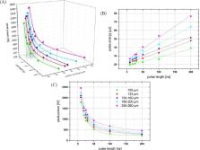
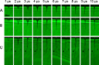
          For more than a decade, living cells and biomaterials (typically hydrogels) are printed via laser-assisted bioprinting. Often, a thin metal layer is applied as laser-absorbing material called dynamic release layer (DRL). This layer is vaporized by focused laser pulses generating vapor pressure that propels forward a coated biomaterial. Different lasers with laser wavelengths from 193 to 1064 nanometer have been used. As a metal DRL gold, silver, or titanium layers have been used. The applied laser pulse durations were usually in the nanosecond range from 1 to 30 ns. In addition, some studies with femtosecond lasers have been published. However, there are no studies on the effect of all these lasers parameters on bioprinting with a metal DRL, and on comparing different wavelengths and pulse durations – except one study comparing 500 femtosecond pulses with 15 ns pulses. In this paper, the effects of laser wavelength (355, 532, and 1064 nm) and laser pulse duration (in the range of 8 to 200 ns) are investigated. Furthermore, the effects of laser pulse energy, intensity, and focal spot size are studied. The printed droplet volume, hydrogel jet velocity, and cell viability are analyzed.

          Related collections

          Most cited references18

          • Record: found
          • Abstract: found
          • Article: not found

          Controlling laser-induced jet formation for bioprinting mesenchymal stem cells with high viability and high resolution.

          Laser-assisted bioprinting is a versatile, non-contact, nozzle-free printing technique which has demonstrated high potential for cell printing with high resolution. Improving cell viability requires determining printing conditions which minimize shear stress for cells within the jet and cell impact at droplet landing. In this context, this study deals with laser-induced jet dynamics to determine conditions from which jets arise with minimum kinetic energies. The transition from a sub-threshold regime to jetting regime has been associated with a geometrical parameter (vertex angle) which can be harnessed to print mesenchymal stem cells with high viability using slow jet conditions. Finally, hydrodynamic jet stability is also studied for higher laser pulse energies which give rise to supersonic but turbulent jets.
            Bookmark
            • Record: found
            • Abstract: found
            • Article: found
            Is Open Access

            Dispensing pico to nanolitre of a natural hydrogel by laser-assisted bioprinting

            Background Laser-assisted bioprinting of multi-cellular replicates in accordance with CAD blueprint may substantially improve our understandings of fundamental aspects of 3 D cell-cell and cell-matrix interactions in vitro. For predictable printing results, a profound knowledge about effects of different processing parameters is essential for realisation of 3 D cell models with well-defined cell densities. Methods Time-resolved imaging of the hydrogel jet dynamics and quantitative assessment of the dependence of printed droplet diameter on the process characteristics were conducted. Results The existence of a counterjet was visualised, proving the bubble collapsing theory for the jet formation. Furthermore, by adjusting the viscosity and height of the applied hydrogel layer in combination with different laser pulse energies, the printing of volumes in the range of 10 to 7000 picolitres was demonstrated. Additionally, the relationship between the viscosity and the layer thickness at different laser pulse energies on the printed droplet volume was identified. Conclusions These findings are essential for the advancement of laser-assisted bioprinting by enabling predictable printing results and the integration of computational methods in the generation of 3 D multi-cellular constructs.
              Bookmark
              • Record: found
              • Abstract: found
              • Article: not found

              PLGA/hydrogel biopapers as a stackable substrate for printing HUVEC networks via BioLP.

              Two major challenges in tissue engineering are mimicking the native cell-cell arrangements of tissues and maintaining viability of three-dimension (3D) tissues thicker than 300 µm. Cell printing and prevascularization of engineered tissues are promising approaches to meet these challenges. However, the printing technologies used in biofabrication must balance the competing parameters of resolution, speed, and volume, which limit the resolution of thicker 3D structures. We suggest that high-resolution conformal printing techniques can be used to print 2D patterns of vascular cells onto biopaper substrates which can then be stacked to form a thicker tissue construct. Towards this end we created 1 cm × 1 cm × 300 µm biopapers to be used as the transferable, stackable substrate for cell printing. 3.6% w/v poly-lactide-co-glycolide was dissolved in chloroform and poured into molds filled with NaCl crystals. The salt was removed with DI water and the scaffolds were dried and loaded with a Collagen Type I or Matrigel. SEM of the biopapers showed extensive porosity and gel loading throughout. Biological laser printing (BioLP) was used to deposit human umbilical vein endothelial cells (HUVEC) in a simple intersecting pattern to the surface of the biopapers. The cells differentiated and stretched to form networks preserving the printed pattern. In a separate experiment to demonstrate "stackability," individual biopapers were randomly seeded with HUVECs and cultured for 1 day. The mechanically stable and viable biopapers were then stacked and cultured for 4 days. Three-dimensional confocal microscopy showed cell infiltration and survival in the compound multilayer constructs. These results demonstrate the feasibility of stackable "biopapers" as a scaffold to build 3D vascularized tissues with a 2D cell-printing technique.
                Bookmark

                Author and article information

                Journal
                Int J Bioprint
                Int J Bioprint
                Whioce Publishing Pte. Ltd.
                International Journal of Bioprinting
                Whioce Publishing Pte. Ltd.
                2424-7723
                2424-8002
                2017
                25 January 2017
                : 3
                : 1
                : 001
                Affiliations
                [1 ]Laser Zentrum Hannover e.V., Nanotechnology Department, Hollerithallee 8, 30419 Hannover, Germany
                [2 ]DeutschesElektronen-Synchrotron (DESY), Notkestraße 85, 22607 Hamburg, Germany
                [3 ]Leibniz Universität Hannover, Institut für Quantenoptik, Welfengarten 1, 30167 Hannover, Germany
                Author notes
                [* ] Correspondence to: Lothar Koch, Laser Zentrum Hannover e.V., Nanotechnology Department, Hollerithallee 8, 30419 Hannover, Germany; Email: l.koch@ 123456lzh.de
                Article
                IJB-3-1-96
                10.18063/IJB.2017.01.001
                7575628
                33094176
                74c2650a-0312-4dd7-aa4e-d12e6acacf09
                Copyright: © 2017 Koch, et al.

                This is an open-access article distributed under the terms of the Attribution-NonCommercial 4.0 International 4.0 (CC BY-NC 4.0), which permits all non-commercial use, distribution, and reproduction in any medium provided the original work is properly cited.

                History
                : 01 October 2016
                : 25 November 2016
                Categories
                Research Article

                bioprinting,laser-assisted bioprinting,laser-induced forward transfer,laser absorption layer,laser parametric study

                Comments

                Comment on this article