3
views
0
recommends
+1 Recommend
0 collections
    0
    shares
      • Record: found
      • Abstract: found
      • Article: found
      Is Open Access

      LncRNA CASC9 Affects Cell Proliferation, Migration, and Invasion of Tongue Squamous cell Carcinoma via Regulating miR-423-5p/SOX12 Axes

      research-article

      Read this article at

      Bookmark
          There is no author summary for this article yet. Authors can add summaries to their articles on ScienceOpen to make them more accessible to a non-specialist audience.

          Abstract

          Introduction

          The incidence of tongue squamous cell carcinoma (TSCC) has increased in recent decades. However, the function of long non-coding RNA (lncRNA) CASC9 in the occurrence and progression of TSCC is unclear. In this work, we attempted to clarify the role of lncRNA CASC9 in determining the phenotype of TSCC cells, and to clarify the underlying mechanisms.

          Methods

          We used qRT-PCR analysis to identify the level of CASC9 mRNA expression in TSCC clinical samples and cell lines. We investigated cell proliferation, and cell migration and invasion of TSCC cells transfected with siCASC9 or siNC using CCK-8 and transwell assays. Bioinformatics analysis and a luciferase reporter assay were employed to predict and verify the target microRNA (miRNA).

          Results

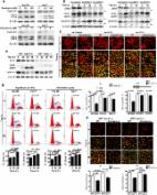
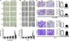
          CASC9 was up-regulated in the TSCC tissues and cells, and predicted a poor prognosis. CASC9 silencing significantly inhibited cell proliferation, migration, and invasion of the TSCC cells compared with the non-targeting control small interfering RNA (siCtrl) treatment. miR-423-5p was predicted as the targeting miRNA of CASC9; this was verified by a luciferase reporter assay. CASC9 expression showed a negative correlation with miR-423-5p expression and a positive correlation with SOX12 expression. The miR-423-5p inhibitor can rescue the carcinogenesis effect of CASC9 on TSCC cells.

          Conclusion

          Our work indicates that CASC9 plays a role in TSCC tumorigenesis; this novel information will improve TSCC molecular targeting therapy.

          Related collections

          Most cited references27

          • Record: found
          • Abstract: found
          • Article: found
          Is Open Access

          Hepatocellular carcinoma–related cyclin D1 is selectively regulated by autophagy degradation system

          Dysfunction of degradation machineries causes cancers, including hepatocellular carcinoma (HCC). Overexpression of cyclin D1 in HCC has been reported. We previously reported that autophagy preferentially recruits and degrades the oncogenic microRNA (miR)‐224 to prevent HCC. Therefore, in the present study, we attempted to clarify whether cyclin D1 is another oncogenic factor selectively regulated by autophagy in HCC tumorigenesis. Initially, we found an inverse correlation between low autophagic activity and high cyclin D1 expression in tumors of 147 HCC patients and three murine models, and these results taken together revealed a correlation with poor overall survival of HCC patients, indicating the importance of these two events in HCC development. We found that increased autophagic activity leads to cyclin D1 ubiquitination and selective recruitment to the autophagosome (AP) mediated by a specific receptor, sequestosome 1 (SQSTM1), followed by fusion with lysosome and degradation. Autophagy‐selective degradation of ubiquitinated cyclin D1 through SQSTM1 was confirmed using cyclin D1/ubiquitin binding site (K33‐238R) and phosphorylation site (T286A) mutants, lentivirus‐mediated silencing autophagy‐related 5 (ATG5), autophagy‐related 7 (ATG7), and Sqstm1 knockout cells. Functional studies revealed that autophagy‐selective degradation of cyclin D1 plays suppressive roles in cell proliferation, colony, and liver tumor formation. Notably, an increase of autophagic activity by pharmacological inducers (amiodarone and rapamycin) significantly suppressed tumor growth in both the orthotopic liver tumor and subcutaneous tumor xenograft models. Our findings provide evidence of the underlying mechanism involved in the regulation of cyclin D1 by selective autophagy to prevent tumor formation. Conclusion: Taken together, our data demonstrate that autophagic degradation machinery and the cell‐cycle regulator, cyclin D1, are linked to HCC tumorigenesis. We believe these findings may be of value in the development of alternative therapeutics for HCC patients. (Hepatology 2018;68:141‐154).
            Bookmark
            • Record: found
            • Abstract: found
            • Article: found
            Is Open Access

            A review of the most promising biomarkers for early diagnosis and prognosis prediction of tongue squamous cell carcinoma

            Background There is a great interest in developing biomarkers to enhance early detection and clinical management of tongue squamous cell carcinoma (TSCC). However, the developmental path towards a clinically valid biomarker remains extremely challenging. Ideally, the initial key step in moving a newly discovered biomarker towards clinical implementation is independent replication. Therefore, the focus of this review is on biomarkers that consistently showed clinical relevance in two or more publications. Methods We searched PubMed database for relevant papers across different TSCC sample sources, i.e., body fluids (saliva, serum/plasma) and tissues. No restriction regarding the date of publication was applied except for immunohistochemistry (IHC); only studies published between 2010 and June 2017 were included. Results The search strategy identified 1429 abstracts, of which 96 papers, examining 150 biomarkers, were eventually included. Of these papers, 66% were exploratory studies evaluating single or a panel of biomarkers in one publication. Ultimately, based on studies that had undergone validation for their clinical relevance in at least two independent studies, we identified 10 promising candidates, consisting of different types of molecules (IL-6, IL-8, and Prolactin in liquid samples; HIF-1α, SOX2, E-cadherin, vimentin, MALAT1, TP53, and NOTCH1 in tissue biopsies) Conclusions Although more exploratory research is needed with newer methods to identify biomarkers for TSCC, rigorous validation of biomarkers that have already shown unbiased assessment in at least two publications should be considered a high priority. Further research on these promising biomarkers or their combination in multi-institutional studies, could provide new possibilities to develop a specific panel for early diagnosis, prognosis, and individualized treatments.
              Bookmark
              • Record: found
              • Abstract: found
              • Article: found
              Is Open Access

              The long noncoding RNA CASC9 regulates migration and invasion in esophageal cancer

              Abstract The objective of the study was to investigate the expression and functions of CASC9 in esophageal squamous cell carcinoma (ESCC). Long noncoding RNAs (lncRNAs) upregulated in ESCC tissues were detected by RNA sequencing. Expression of CASC9 was determined from clinical samples and cell lines by qRT‐PCR. The effects of CASC9 knockdown on migration and invasion were evaluated by wound healing assay, cell migration and invasion assays in vitro. We found that the lncRNA, CASC9, was markedly upregulated in ESCC tissues. Furthermore, knockdown of CASC9 significantly suppressed cell migration and invasion in vitro. Furthermore, enhanced CASC9 expression level was correlated with differentiation. The results indicated that CASC9 is significantly upregulated in ESCC tissues and may represent a new marker of poor prognosis and a potential therapeutic target for esophageal cancer intervention.
                Bookmark

                Author and article information

                Journal
                Cancer Manag Res
                Cancer Manag Res
                CMAR
                cancmanres
                Cancer Management and Research
                Dove
                1179-1322
                14 January 2020
                2020
                : 12
                : 277-287
                Affiliations
                [1 ]Department of Oral and Maxillofacial Surgery, Nanjing Stomatological Hospital, Medical School of Nanjing University , Nanjing, People’s Republic of China
                [2 ]Oncology Department, The Second Hospital of Nanjing , Nanjing, People’s Republic of China
                Author notes
                Correspondence: Ying Zhang Oncology Department, The Second Hospital of Nanjing , No. 1-1 Zhongfu Road, Nanjing210003, People’s Republic of ChinaTel +86-025-83626473 Email yingzhang2018026@126.com
                Article
                220351
                10.2147/CMAR.S220351
                6969678
                7630f2e3-fefc-42c5-87ec-a48127a84781
                © 2020 Chen et al.

                This work is published and licensed by Dove Medical Press Limited. The full terms of this license are available at https://www.dovepress.com/terms.php and incorporate the Creative Commons Attribution – Non Commercial (unported, v3.0) License ( http://creativecommons.org/licenses/by-nc/3.0/). By accessing the work you hereby accept the Terms. Non-commercial uses of the work are permitted without any further permission from Dove Medical Press Limited, provided the work is properly attributed. For permission for commercial use of this work, please see paragraphs 4.2 and 5 of our Terms ( https://www.dovepress.com/terms.php).

                History
                : 21 June 2019
                : 19 November 2019
                Page count
                Figures: 5, Tables: 1, References: 29, Pages: 11
                Categories
                Original Research

                Oncology & Radiotherapy
                casc9,tongue squamous cell carcinoma,mir-423-5p,proliferation,invasion,migration

                Comments

                Comment on this article