14
views
0
recommends
+1 Recommend
0 collections
    0
    shares
      • Record: found
      • Abstract: found
      • Article: not found

      Stem Cell Imaging: Tools to Improve Cell Delivery and Viability

      review-article
      , *
      Stem Cells International
      Hindawi Publishing Corporation

      Read this article at

      ScienceOpenPublisherPMC
      Bookmark
          There is no author summary for this article yet. Authors can add summaries to their articles on ScienceOpen to make them more accessible to a non-specialist audience.

          Abstract

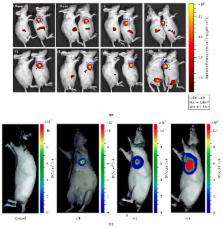
          Stem cell therapy (SCT) has shown very promising preclinical results in a variety of regenerative medicine applications. Nevertheless, the complete utility of this technology remains unrealized. Imaging is a potent tool used in multiple stages of SCT and this review describes the role that imaging plays in cell harvest, cell purification, and cell implantation, as well as a discussion of how imaging can be used to assess outcome in SCT. We close with some perspective on potential growth in the field.

          Related collections

          Most cited references118

          • Record: found
          • Abstract: found
          • Article: not found

          A molecular imaging primer: modalities, imaging agents, and applications.

          Molecular imaging is revolutionizing the way we study the inner workings of the human body, diagnose diseases, approach drug design, and assess therapies. The field as a whole is making possible the visualization of complex biochemical processes involved in normal physiology and disease states, in real time, in living cells, tissues, and intact subjects. In this review, we focus specifically on molecular imaging of intact living subjects. We provide a basic primer for those who are new to molecular imaging, and a resource for those involved in the field. We begin by describing classical molecular imaging techniques together with their key strengths and limitations, after which we introduce some of the latest emerging imaging modalities. We provide an overview of the main classes of molecular imaging agents (i.e., small molecules, peptides, aptamers, engineered proteins, and nanoparticles) and cite examples of how molecular imaging is being applied in oncology, neuroscience, cardiology, gene therapy, cell tracking, and theranostics (therapy combined with diagnostics). A step-by-step guide to answering biological and/or clinical questions using the tools of molecular imaging is also provided. We conclude by discussing the grand challenges of the field, its future directions, and enormous potential for further impacting how we approach research and medicine.
            Bookmark
            • Record: found
            • Abstract: found
            • Article: not found

            Safety and immunological effects of mesenchymal stem cell transplantation in patients with multiple sclerosis and amyotrophic lateral sclerosis.

            To evaluate the feasibility, safety, and immunological effects of intrathecal and intravenous administration of autologous mesenchymal stem cells (MSCs) (also called mesenchymal stromal cells) in patients with multiple sclerosis (MS) and amyotrophic lateral sclerosis (ALS). A phase 1/2 open-safety clinical trial. Patients  Fifteen patients with MS (mean [SD] Expanded Disability Status Scale [EDSS] score, 6.7 [1.0]) and 19 with ALS (mean [SD] Amyotrophic Lateral Sclerosis Functional Rating Scale [ALSFRS] score, 20.8 [8.0]) were enrolled. Intervention  After culture, a mean (SD) of 63.2 × 10(6) (2.5 × 10(6)) MSCs was injected intrathecally (n = 34) and intravenously (n = 14). In 9 cases, MSCs were magnetically labeled with the superparamagnetic iron oxide ferumoxides (Feridex). The main outcome measure was the recording of side effects. Follow-up (≤25 months) included adverse events evaluation, neurological disability assessment by means of the EDSS, magnetic resonance imaging to exclude unexpected pathologies and track the labeled stem cells, and immunological tests to assess the short-term immunomodulatory effects of MSC transplantation. Twenty-one patients had injection-related adverse effects consisting of transient fever, and 15 reported headache. No major adverse effects were reported during follow-up. The mean ALSFRS score remained stable during the first 6 months of observation, whereas the mean (SD) EDSS score improved from 6.7 (1.0) to 5.9 (1.6). Magnetic resonance imaging visualized the MSCs in the occipital horns of the ventricles, indicating the possible migration of ferumoxides-labeled cells in the meninges, subarachnoid space, and spinal cord. Immunological analysis revealed an increase in the proportion of CD4(+)CD25(+) regulatory T cells, a decrease in the proliferative responses of lymphocytes, and the expression of CD40(+), CD83(+), CD86(+), and HLA-DR on myeloid dendritic cells at 24 hours after MSC transplantation. Transplantation of MSCs in patients with MS and ALS is a clinically feasible and relatively safe procedure and induces immediate immunomodulatory effects. Trial Registration  clinicaltrials.gov Identifier: NCT00781872.
              Bookmark
              • Record: found
              • Abstract: found
              • Article: not found

              Identification of ROS using oxidized DCFDA and flow-cytometry.

              Cells constantly generate reactive oxygen species (ROS) during aerobic metabolism. The ROS generation plays an important protective and functional role in the immune system. The cell is armed with a powerful antioxidant defense system to combat excessive production of ROS. Oxidative stress occurs in cells when the generation of ROS overwhelms the cells' natural antioxidant defenses. ROS and the oxidative damage are thought to play an important role in many human diseases including cancer, atherosclerosis, other neurodegenerative diseases and diabetes. Thus, establishing their precise role requires the ability to measure ROS accurately and the oxidative damage that they cause. There are many methods for measuring free radical production in cells. The most straightforward techniques use cell permeable fluorescent and chemiluminescent probes. 2'-7'-Dichlorodihydrofluorescein diacetate (DCFH-DA) is one of the most widely used techniques for directly measuring the redox state of a cell. It has several advantages over other techniques developed. It is very easy to use, extremely sensitive to changes in the redox state of a cell, inexpensive and can be used to follow changes in ROS over time.
                Bookmark

                Author and article information

                Journal
                Stem Cells Int
                Stem Cells Int
                SCI
                Stem Cells International
                Hindawi Publishing Corporation
                1687-966X
                1687-9678
                2016
                6 January 2016
                : 2016
                : 9240652
                Affiliations
                Department of Nanoengineering, University of California, San Diego, 9500 Gilman Drive, La Jolla, CA 92093-0448, USA
                Author notes
                *Jesse V. Jokerst: jjokerst@ 123456ucsd.edu

                Academic Editor: Olivier Gheysens

                Article
                10.1155/2016/9240652
                4736428
                26880997
                78ee39e3-9970-410b-b0bb-bc00924f97bf
                Copyright © 2016 J. Wang and J. V. Jokerst.

                This is an open access article distributed under the Creative Commons Attribution License, which permits unrestricted use, distribution, and reproduction in any medium, provided the original work is properly cited.

                History
                : 8 July 2015
                : 6 October 2015
                : 15 October 2015
                Categories
                Review Article

                Molecular medicine
                Molecular medicine

                Comments

                Comment on this article