2
views
0
recommends
+1 Recommend
0 collections
    0
    shares
      • Record: found
      • Abstract: found
      • Article: found
      Is Open Access

      Analysis of 3-Dimensional Arch Anatomy, Vascular Flow, and Postnatal Outcome in Cases of Suspected Coarctation of the Aorta Using Fetal Cardiac Magnetic Resonance Imaging

      research-article

      Read this article at

      Bookmark
          There is no author summary for this article yet. Authors can add summaries to their articles on ScienceOpen to make them more accessible to a non-specialist audience.

          Abstract

          Supplemental Digital Content is available in the text.

          Background:

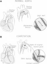
          Identifying fetuses at risk of severe neonatal coarctation of the aorta (CoA) can be lifesaving but is notoriously challenging in clinical practice with a high rate of false positives. Novel fetal 3-dimensional and phase-contrast magnetic resonance imaging (MRI) offers an unprecedented means of assessing the human fetal cardiovascular system before birth. We performed detailed MRI assessment of fetal vascular morphology and flows in a cohort of fetuses with suspected CoA, correlated with the need for postnatal intervention.

          Methods:

          Women carrying a fetus with suspected CoA on echocardiography were referred for MRI assessment between 26 and 36 weeks of gestation, including high-resolution motion-corrected 3-dimensional volumes of the fetal heart and phase-contrast flow sequences gated with metric optimized gating. The relationship between aortic geometry and vascular flows was then analyzed and compared with postnatal outcome.

          Results:

          Seventy-two patients (51 with suspected fetal CoA and 21 healthy controls) underwent fetal MRI with motion-corrected 3-dimensional vascular reconstructions. Vascular flow measurements from phase-contrast sequences were available in 53 patients. In the CoA group, 25 of 51 (49%) required surgical repair of coarctation after birth; the remaining 26 of 51 (51%) were discharged without neonatal intervention. Reduced blood flow in the fetal ascending aorta and at the aortic isthmus was associated with increasing angulation ( P=0.005) and proximal displacement ( P=0.006) of the isthmus and was seen in both true positive and false positive cases. A multivariate logistic regression model including aortic flow and isthmal displacement explained 78% of the variation in outcome and correctly predicted the need for intervention in 93% of cases.

          Conclusions:

          Reduced blood flow though the left heart is associated with important configurational changes at the aortic isthmus in fetal life, predisposing to CoA when the arterial duct closes after birth. Novel fetal MRI techniques may have a role in both understanding and accurately predicting severe neonatal CoA.

          Related collections

          Most cited references43

          • Record: found
          • Abstract: found
          • Article: not found

          Aortic dilation in bicuspid aortic valve disease: flow pattern is a major contributor and differs with valve fusion type.

          Ascending aortic dilation is important in bicuspid aortic valve (BAV) disease, with increased risk of aortic dissection. We used cardiovascular MR to understand the pathophysiology better by examining the links between 3-dimensional flow abnormalities, aortic function, and aortic dilation. A total of 142 subjects underwent cardiovascular MR (mean age, 40 years; 95 with BAV, 47 healthy volunteers). Patients with BAV had predominantly abnormal right-handed helical flow in the ascending aorta, larger ascending aortas (18.3±3.3 versus 15.2±2.2 mm/m²; P<0.001), and higher rotational (helical) flow (31.7±15.8 versus 2.9±3.9 mm²/s; P<0.001), systolic flow angle (23.1°±12.5° versus 7.0°±4.6°; P<0.001), and systolic wall shear stress (0.85±0.28 versus 0.59±0.17 N/m²; P<0.001) compared with healthy volunteers. BAV with right-handed flow and right-non coronary cusp fusion (n=31) showed more severe flow abnormalities (rotational flow, 38.5±16.5 versus 27.8±12.4 mm²/s; P<0.001; systolic flow angle, 29.4°±10.9° versus 19.4°±11.4°; P<0.001; in-plane wall shear stress, 0.64±0.23 versus 0.47±0.22 N/m²; P<0.001) and larger aortas (19.5±3.4 versus 17.5±3.1 mm/m²; P<0.05) than right-left cusp fusion (n=55). Patients with BAV with normal flow patterns had similar aortic dimensions and wall shear stress to healthy volunteers and younger patients with BAV showed abnormal flow patterns but no aortic dilation, both further supporting the importance of flow pattern in the pathogenesis of aortic dilation. Aortic function measures (distensibility, aortic strain, and pulse wave velocity) were similar across all groups. Flow abnormalities may be a major contributor to aortic dilation in BAV. Fusion type affects the severity of flow abnormalities and may allow better risk prediction and selection of patients for earlier surgical intervention.
            Bookmark
            • Record: found
            • Abstract: found
            • Article: not found

            Diagnosis, imaging and clinical management of aortic coarctation.

            Coarctation of the aorta (CoA ) is a well-known congenital heart disease (CHD) , which is often associated with several other cardiac and vascular anomalies, such as bicuspid aortic valve (BAV), ventricular septal defect, patent ductus arteriosus and aortic arch hypoplasia. Despite echocardiographic screening, prenatal diagnosis of C o A remains difficult. Most patients with CoA present in infancy with absent, delayed or reduced femoral pulses, a supine arm-leg blood pressure gradient (> 20 mm Hg), or a murmur due to rapid blood flow across the CoA or associated lesions (BAV). Transthoracic echocardiography is the primary imaging modality for suspected CoA. However, cardiac magnetic resonance imaging is the preferred advanced imaging modality for non-invasive diagnosis and follow-up of CoA. Adequate and timely diagnosis of CoA is crucial for good prognosis, as early treatment is associated with lower risks of long-term morbidity and mortality. Numerous surgical and transcatheter treatment strategies have been reported for CoA. Surgical resection is the treatment of choice in neonates, infants and young children. In older children (> 25 kg) and adults, transcatheter treatment is the treatment of choice. In the current era, patients with CoA continue to have a reduced life expectancy and an increased risk of cardiovascular sequelae later in life, despite adequate relief of the aortic stenosis. Intensive and adequate follow-up of the left ventricular function, valvular function, blood pressure and the anatomy of the heart and the aorta are , therefore, critical in the management of CoA. This review provides an overview of the current state-of-the-art clinical diagnosis, diagnostic imaging algori thms, treatment and follow-up of patients with CoA.
              Bookmark
              • Record: found
              • Abstract: found
              • Article: not found

              Prenatal diagnosis of coarctation of the aorta improves survival and reduces morbidity.

              To investigate whether antenatal diagnosis of coarctation of the aorta results in reduced mortality and improved preoperative haemodynamic stability compared with postnatal diagnosis. Retrospective review of all cases of coarctation of the aorta presenting to a tertiary fetal and neonatal cardiology service from January 1994 to December 1998. Prenatal, postnatal, and necropsy records were reviewed to determine survival in the two groups. Markers of preoperative illness severity were recorded, including presence of femoral pulse, collapse, left ventricular function, ductal patency on echocardiography, coagulation status, duration of intensive care unit and total hospital stay, heart rate, respiratory rate, plasma creatinine, plasma potassium, and right upper limb blood pressure. A univarate and multivariate analysis was conducted on all variables and a cumulative score was created and subjected to logistic regression analysis. Both collapse and death were more common in the postnatally diagnosed group (p < 0.05). Femoral pulses were more likely to be palpable and there was echocardiographic evidence of duct patency in the antenatally diagnosed infants (p < 0.001 and p < 0.05, respectively). An increased respiratory rate was associated with postnatal presentation (p < 0.05). Infants with haemodynamic instability preoperatively were more likely to have been diagnosed postnatally (p < 0.01). Antenatal diagnosis of coarctation of the aorta is associated with improved survival and preoperative clinical condition.
                Bookmark

                Author and article information

                Contributors
                Journal
                Circ Cardiovasc Imaging
                Circ Cardiovasc Imaging
                HCI
                Circulation. Cardiovascular Imaging
                Lippincott Williams & Wilkins (Hagerstown, MD )
                1941-9651
                1942-0080
                30 June 2021
                July 2021
                : 14
                : 7
                : e012411
                Affiliations
                [1 ]School of Imaging Sciences and Biomedical Engineering, King’s College London, United Kingdom (D.F.A.L., M.P.M.v.P., K.P., J.S., J.F.P.v.A., T.R., A.S., M.R., J.H., R.R.).
                [2 ]Department of Congenital Heart Disease, Evelina London Children’s Hospital, United Kingdom (D.F.A.L., K.P., T.V.V., V.Z., M.C., O.M., G.S., J.M.S., R.R.).
                Author notes
                Correspondence to: Reza Razavi, MD, Evelina London Children’s Hospital, Room 8.11, 8th Floor, Melbourne House, London WC2B 4LL, England. Email reza.razavi@ 123456kcl.ac.uk
                Article
                00002
                10.1161/CIRCIMAGING.121.012411
                8300852
                34187165
                7905e540-1f5c-4f09-8685-0526104900ec
                © 2021 The Authors.

                Circulation: Cardiovascular Imaging is published on behalf of the American Heart Association, Inc., by Wolters Kluwer Health, Inc. This is an open access article under the terms of the Creative Commons Attribution License, which permits use, distribution, and reproduction in any medium, provided that the original work is properly cited.

                History
                : 7 January 2021
                : 14 April 2021
                Categories
                10015
                10067
                10093
                10129
                10132
                Original Articles
                Custom metadata
                TRUE
                T

                biomarkers,heart defects, congenital,magnetic resonance imaging,pediatrics,prognosis

                Comments

                Comment on this article