23
views
0
recommends
+1 Recommend
0 collections
    0
    shares
      • Record: found
      • Abstract: found
      • Article: found
      Is Open Access

      Metabolic Aspects of Neuron-Oligodendrocyte-Astrocyte Interactions

      review-article

      Read this article at

      Bookmark
          There is no author summary for this article yet. Authors can add summaries to their articles on ScienceOpen to make them more accessible to a non-specialist audience.

          Abstract

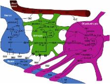
          Whereas astrocytes have been in the limelight of scientific interest in brain energy metabolism for a while, oligodendrocytes are still waiting for a place on the metabolic stage. We propose to term the interaction of oligodendrocytes with astrocytes and neurons: NOA (neuron–oligodendrocyte–astrocyte) interactions. One of the reasons to find out more about metabolic interactions between oligodendrocytes, neurons, and astrocytes is to establish markers of healthy oligodendrocyte metabolism that could be used for the diagnosis and assessment of white matter disease. The vesicular release of glutamate in the white matter has received considerable attention in the past. Oligodendrocyte lineage cells express glutamate receptors and glutamate toxicity has been implicated in diseases affecting oligodendrocytes such as hypoxic-ischaemic encephalopathy, inflammatory diseases and trauma. As oligodendrocyte precursor cells vividly react to injury it is also important to establish whether cells recruited into damaged areas are able to regenerate lost myelin sheaths or whether astrocytic scarring occurs. It is therefore important to consider metabolic aspects of astrocytes and oligodendrocytes separately. The present review summarizes the limited evidence available on metabolic cycles in oligodendrocytes and so hopes to stimulate further research interests in this important field.

          Related collections

          Most cited references50

          • Record: found
          • Abstract: found
          • Article: not found

          NMDA receptors are expressed in oligodendrocytes and activated in ischaemia.

          Glutamate-mediated damage to oligodendrocytes contributes to mental or physical impairment in periventricular leukomalacia (pre- or perinatal white matter injury leading to cerebral palsy), spinal cord injury, multiple sclerosis and stroke. Unlike neurons, white matter oligodendrocytes reportedly lack NMDA (N-methyl-d-aspartate) receptors. It is believed that glutamate damages oligodendrocytes, especially their precursor cells, by acting on calcium-permeable AMPA (alpha-amino-3-hydroxy-5-methyl-4-isoxazole propionic acid)/kainate receptors alone or by reversing cystine-glutamate exchange and depriving cells of antioxidant protection. Here we show that precursor, immature and mature oligodendrocytes in the white matter of the cerebellum and corpus callosum exhibit NMDA-evoked currents, mediated by receptors that are blocked only weakly by Mg2+ and that may contain NR1, NR2C and NR3 NMDA receptor subunits. NMDA receptors are present in the myelinating processes of oligodendrocytes, where the small intracellular space could lead to a large rise in intracellular ion concentration in response to NMDA receptor activation. Simulating ischaemia led to development of an inward current in oligodendrocytes, which was partly mediated by NMDA receptors. These results point to NMDA receptors of unusual subunit composition as a potential therapeutic target for preventing white matter damage in a variety of diseases.
            Bookmark
            • Record: found
            • Abstract: found
            • Article: not found

            Vesicular glutamate release from axons in white matter.

            Vesicular release of neurotransmitter is the universal output signal of neurons in the brain. It is generally believed that fast transmitter release is restricted to nerve terminals that contact postsynaptic cells in the gray matter. Here we show in the rat brain that the neurotransmitter glutamate is also released at discrete sites along axons in white matter in the absence of neurons and nerve terminals. The propagation of single action potentials along axons leads to rapid vesicular release of glutamate, which is detected by ionotropic glutamate receptors on local oligodendrocyte precursor cells. Axonal release of glutamate is reliable, involves highly localized calcium microdomain signaling and is strongly calcium cooperative, similar to vesicle fusion at synapses. This axonal transmitter release represents a widespread mechanism for high-fidelity, activity-dependent signaling at the axon-glia interface in white matter.
              Bookmark
              • Record: found
              • Abstract: found
              • Article: not found

              Vesicular release of glutamate from unmyelinated axons in white matter.

              Directed fusion of transmitter-laden vesicles enables rapid intercellular signaling in the central nervous system and occurs at synapses within gray matter. Here we show that action potentials also induce the release of glutamate from axons in the corpus callosum, a white matter region responsible for interhemispheric communication. Callosal axons release glutamate by vesicular fusion, which induces quantal AMPA receptor-mediated currents in NG2(+) glial progenitors at anatomically distinct axo-glial synaptic junctions. Glutamate release from axons was facilitated by repetitive stimulation and could be inhibited through activation of metabotropic autoreceptors. Although NG2(+) cells form associations with nodes of Ranvier in white matter, measurements of conduction velocity indicated that unmyelinated fibers are responsible for glutamatergic signaling with NG2(+) glia. This activity-dependent secretion of glutamate was prevalent in the developing and mature mouse corpus callosum, indicating that axons within white matter both conduct action potentials and engage in rapid neuron-glia communication.
                Bookmark

                Author and article information

                Journal
                Front Endocrinol (Lausanne)
                Front Endocrinol (Lausanne)
                Front. Endocrinol.
                Frontiers in Endocrinology
                Frontiers Media S.A.
                1664-2392
                13 May 2013
                2013
                : 4
                : 54
                Affiliations
                [1] 1Anne McLaren Laboratory for Regenerative Medicine, Wellcome Trust and Medical Research Council Cambridge Stem Cell Institute, Department of Clinical Neurosciences, University of Cambridge Cambridge, UK
                [2] 2Department of Neuroscience, Faculty of Medicine, Norwegian University of Science and Technology Trondheim, Norway
                Author notes

                Edited by: Leif Hertz, Medical University of China, China

                Reviewed by: Anne Hélène Duittoz, Université de Tours, France; Leif Hertz, Medical University of China, China; Jacques De Keyser, Universitair Ziekenhuis Vrije Universiteit Brussel, Belgium

                *Correspondence: Ursula Sonnewald, Department of Neuroscience, Faculty of Medicine, Norwegian University of Science and Technology, PO Box 8905, MTFS, 7491 Trondheim, Norway. e-mail: ursula.sonnewald@ 123456ntnu.no

                This article was submitted to Frontiers in Cellular Endocrinology, a specialty of Frontiers in Endocrinology.

                Article
                10.3389/fendo.2013.00054
                3651962
                23717302
                79247a71-78cb-4297-a02a-4aaa8ece1c47
                Copyright © 2013 Amaral, Meisingset, Kotter and Sonnewald.

                This is an open-access article distributed under the terms of the Creative Commons Attribution License, which permits use, distribution and reproduction in other forums, provided the original authors and source are credited and subject to any copyright notices concerning any third-party graphics etc.

                History
                : 12 March 2013
                : 24 April 2013
                Page count
                Figures: 1, Tables: 0, Equations: 0, References: 59, Pages: 5, Words: 4611
                Categories
                Endocrinology
                Mini Review

                Endocrinology & Diabetes
                energy metabolism,glucose,gray matter,white matter,pyruvate carboxylation,glycolysis,lactate,monocarboxylate transporters

                Comments

                Comment on this article