63
views
0
recommends
+1 Recommend
0 collections
    0
    shares
      • Record: found
      • Abstract: found
      • Article: found
      Is Open Access

      Acrolein Induces Endoplasmic Reticulum Stress and Causes Airspace Enlargement

      research-article

      Read this article at

      Bookmark
          There is no author summary for this article yet. Authors can add summaries to their articles on ScienceOpen to make them more accessible to a non-specialist audience.

          Abstract

          Background

          Given the relative abundance and toxic potential of acrolein in inhaled cigarette smoke, it is surprising how little is known about the pulmonary and systemic effects of acrolein. Here we test the hypothesis whether systemic administration of acrolein could cause endoplasmic reticulum (ER) stress, and lung cell apoptosis, leading to the enlargement of the alveolar air spaces in rats.

          Methods

          Acute and chronic effects of intraperitoneally administered acrolein were tested. Mean alveolar airspace area was measured by using light microscopy and imaging system software. TUNEL staining and immunohistochemistry (IHC) for active caspase 3 and Western blot analysis for active caspase 3, and caspase 12 were performed to detect apoptosis. The ER-stress related gene expression in the lungs was determined by Quantitative real-time PCR analysis. Acrolein-protein adducts in the lung tissue were detected by IHC.

          Results

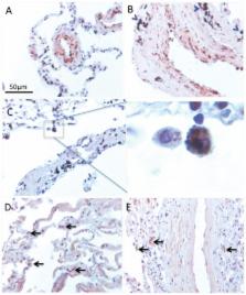
          Acute administration of acrolein caused a significant elevation of activated caspase 3, upregulation of VEGF expression and induced ER stress proteins in the lung tissue. The chronic administration of acrolein in rats led to emphysematous lung tissue remodeling. TUNEL staining and IHC for cleaved caspase 3 showed a large number of apoptotic septal cells in the acrolein-treated rat lungs. Chronic acrolein administration cause the endoplasmic reticulum stress response manifested by significant upregulation of ATF4, CHOP and GADd34 expression. In smokers with COPD there was a considerable accumulation of acrolein-protein adducts in the inflammatory, airway and vascular cells.

          Conclusions

          Systemic administration of acrolein causes endoplasmic reticulum stress response, lung cell apoptosis, and chronic administration leads to the enlargement of the alveolar air spaces and emphysema in rats. The substantial accumulation of acrolein-protein adducts in the lungs of COPD patients suggest a role of acrolein in the pathogenesis of emphysema.

          Related collections

          Most cited references50

          • Record: found
          • Abstract: not found
          • Article: not found

          Indoor air quality and health

          A.P. Jones (1999)
            Bookmark
            • Record: found
            • Abstract: found
            • Article: not found

            Acrolein: sources, metabolism, and biomolecular interactions relevant to human health and disease.

            Acrolein (2-propenal) is ubiquitously present in (cooked) foods and in the environment. It is formed from carbohydrates, vegetable oils and animal fats, amino acids during heating of foods, and by combustion of petroleum fuels and biodiesel. Chemical reactions responsible for release of acrolein include heat-induced dehydration of glycerol, retro-aldol cleavage of dehydrated carbohydrates, lipid peroxidation of polyunsaturated fatty acids, and Strecker degradation of methionine and threonine. Smoking of tobacco products equals or exceeds the total human exposure to acrolein from all other sources. The main endogenous sources of acrolein are myeloperoxidase-mediated degradation of threonine and amine oxidase-mediated degradation of spermine and spermidine, which may constitute a significant source of acrolein in situations of oxidative stress and inflammation. Acrolein is metabolized by conjugation with glutathione and excreted in the urine as mercapturic acid metabolites. Acrolein forms Michael adducts with ascorbic acid in vitro, but the biological relevance of this reaction is not clear. The biological effects of acrolein are a consequence of its reactivity towards biological nucleophiles such as guanine in DNA and cysteine, lysine, histidine, and arginine residues in critical regions of nuclear factors, proteases, and other proteins. Acrolein adduction disrupts the function of these biomacromolecules which may result in mutations, altered gene transcription, and modulation of apoptosis.
              Bookmark
              • Record: found
              • Abstract: found
              • Article: not found

              Endothelial cell death and decreased expression of vascular endothelial growth factor and vascular endothelial growth factor receptor 2 in emphysema.

              Emphysema due to cigarette smoking is characterized by a loss of alveolar structures. We hypothesize that the disappearance of alveoli involves apoptosis of septal endothelial cells and a decreased expression of lung vascular endothelial growth factor (VEGF) and its receptor 2 (VEGF R2). By terminal transferase dUTP nick end labeling (TUNEL) in combination with immunohistochemistry, we found that the number of TUNEL+ septal epithelial and endothelial cells/lung tissue nucleic acid (microg) was increased in the alveolar septa of emphysema lungs (14.2 +/- 2.0/microg, n = 6) when compared with normal lungs (6.8 +/- 1.3/microg, n = 7) (p < 0.01) and with primary pulmonary hypertensive lungs (2.3 +/- 0.8/microg, n = 5) (p < 0.001). The cell death events were not significantly different between healthy nonsmoker (7.4 +/- 1.9/microg) and smoker (5.7 +/- 0.7/microg) control subjects. The TUNEL results were confirmed by single-stranded DNA and active caspase-3 immunohistochemistry, and by DNA ligation assay. Emphysema lungs (n = 12) had increased levels of oligonucleosomal-length DNA fragmentation when compared with normal lungs (n = 11). VEGF, VEGF R2 protein, and mRNA expression were significantly reduced in emphysema. We propose that epithelial and endothelial alveolar septal death due to a decrease of endothelial cell maintenance factors may be part of the pathogenesis of emphysema.
                Bookmark

                Author and article information

                Contributors
                Role: Editor
                Journal
                PLoS One
                PLoS ONE
                plos
                plosone
                PLoS ONE
                Public Library of Science (San Francisco, USA )
                1932-6203
                2012
                31 May 2012
                : 7
                : 5
                : e38038
                Affiliations
                [1 ]Pulmonary and Critical Care Medicine Division, The Victoria Johnson Laboratory for Obstructive Lung Disease Research, Virginia Commonwealth University, Richmond, Virginia, United States of America
                [2 ]First Department of Internal Medicine, Sinshu University School of Medicine, Matsumoto, Japan
                [3 ]Division of Pulmonary Sciences and Critical Care Medicine, University of Colorado School of Medicine, Aurora, Colorado, United States of America
                University of Giessen Lung Center, Germany
                Author notes

                Conceived and designed the experiments: YK LTS NFV. Performed the experiments: YK LTS MH RN DK. Analyzed the data: YK LTS RN. Contributed reagents/materials/analysis tools: NFV LTS. Wrote the paper: YK LTS NFV.

                Article
                PONE-D-11-24831
                10.1371/journal.pone.0038038
                3364999
                22675432
                7a0b3a22-91d2-4630-a78a-f4f7632e94ab
                Kitaguchi et al. This is an open-access article distributed under the terms of the Creative Commons Attribution License, which permits unrestricted use, distribution, and reproduction in any medium, provided the original author and source are credited.
                History
                : 12 December 2011
                : 30 April 2012
                Page count
                Pages: 10
                Categories
                Research Article
                Biology
                Biochemistry
                Histology
                Immunology
                Immunologic Techniques
                Immunohistochemical Analysis
                Molecular Cell Biology
                Signal Transduction
                Signaling Cascades
                Stress Signaling Cascade
                Cellular Stress Responses
                Medicine
                Cardiovascular
                Clinical Immunology
                Immunologic Techniques
                Immunohistochemical Analysis
                Non-Clinical Medicine
                Pulmonology
                Smoking Related Disorders

                Uncategorized
                Uncategorized

                Comments

                Comment on this article