56
views
0
recommends
+1 Recommend
0 collections
    0
    shares
      • Record: found
      • Abstract: found
      • Article: found
      Is Open Access

      A Non-invasive Platform for Functional Characterization of Stem-Cell-Derived Cardiomyocytes with Applications in Cardiotoxicity Testing

      research-article

      Read this article at

      Bookmark
          There is no author summary for this article yet. Authors can add summaries to their articles on ScienceOpen to make them more accessible to a non-specialist audience.

          Summary

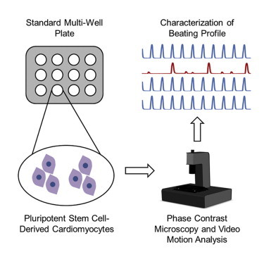
          We present a non-invasive method to characterize the function of pluripotent stem-cell-derived cardiomyocytes based on video microscopy and image analysis. The platform, called Pulse, generates automated measurements of beating frequency, beat duration, amplitude, and beat-to-beat variation based on motion analysis of phase-contrast images captured at a fast frame rate. Using Pulse, we demonstrate recapitulation of drug effects in stem-cell-derived cardiomyocytes without the use of exogenous labels and show that our platform can be used for high-throughput cardiotoxicity drug screening and studying physiologically relevant phenotypes.

          Graphical Abstract

          Highlights

          • Non-invasive characterization of cardiomyocytes using video motion analysis

          • Assessment of single-cell, monolayer, or tissue cardiomyocytes in standard plates

          • Drug screening by a label-free, contact-free imaging assay

          Abstract

          In this article, Maddah and colleagues present a non-invasive method to characterize the function of stem-cell-derived cardiomyocytes based on video microscopy and image analysis. They demonstrate automated measurements of beating frequency, duration, amplitude, and variation and show that the platform can be used for cardiotoxicity drug screening and studying physiologically relevant phenotypes.

          Related collections

          Most cited references18

          • Record: found
          • Abstract: found
          • Article: not found

          Cardiomyocytes can be generated from marrow stromal cells in vitro.

          We have isolated a cardiomyogenic cell line (CMG) from murine bone marrow stromal cells. Stromal cells were immortalized, treated with 5-azacytidine, and spontaneously beating cells were repeatedly screened. The cells showed a fibroblast-like morphology, but the morphology changed after 5-azacytidine treatment in approximately 30% of the cells; they connected with adjoining cells after one week, formed myotube-like structures, began spontaneously beating after two weeks, and beat synchronously after three weeks. They expressed atrial natriuretic peptide and brain natriuretic peptide and were stained with anti-myosin, anti-desmin, and anti-actinin antibodies. Electron microscopy revealed a cardiomyocyte-like ultrastructure, including typical sarcomeres, a centrally positioned nucleus, and atrial granules. These cells had several types of action potentials, such as sinus node-like and ventricular cell-like action potentials. All cells had a long action potential duration or plateau, a relatively shallow resting membrane potential, and a pacemaker-like late diastolic slow depolarization. Analysis of the isoform of contractile protein genes, such as myosin heavy chain, myosin light chain, and alpha-actin, indicated that their muscle phenotype was similar to that of fetal ventricular cardiomyocytes. These cells expressed Nkx2.5/Csx, GATA4, TEF-1, and MEF-2C mRNA before 5-azacytidine treatment and expressed MEF-2A and MEF-2D after treatment. This new cell line provides a powerful model for the study of cardiomyocyte differentiation.
            Bookmark
            • Record: found
            • Abstract: found
            • Article: not found

            Patient-specific induced pluripotent stem cells as a model for familial dilated cardiomyopathy.

            Characterized by ventricular dilatation, systolic dysfunction, and progressive heart failure, dilated cardiomyopathy (DCM) is the most common form of cardiomyopathy in patients. DCM is the most common diagnosis leading to heart transplantation and places a significant burden on healthcare worldwide. The advent of induced pluripotent stem cells (iPSCs) offers an exceptional opportunity for creating disease-specific cellular models, investigating underlying mechanisms, and optimizing therapy. Here, we generated cardiomyocytes from iPSCs derived from patients in a DCM family carrying a point mutation (R173W) in the gene encoding sarcomeric protein cardiac troponin T. Compared to control healthy individuals in the same family cohort, cardiomyocytes derived from iPSCs from DCM patients exhibited altered regulation of calcium ion (Ca(2+)), decreased contractility, and abnormal distribution of sarcomeric α-actinin. When stimulated with a β-adrenergic agonist, DCM iPSC-derived cardiomyocytes showed characteristics of cellular stress such as reduced beating rates, compromised contraction, and a greater number of cells with abnormal sarcomeric α-actinin distribution. Treatment with β-adrenergic blockers or overexpression of sarcoplasmic reticulum Ca(2+) adenosine triphosphatase (Serca2a) improved the function of iPSC-derived cardiomyocytes from DCM patients. Thus, iPSC-derived cardiomyocytes from DCM patients recapitulate to some extent the morphological and functional phenotypes of DCM and may serve as a useful platform for exploring disease mechanisms and for drug screening.
              Bookmark
              • Record: found
              • Abstract: found
              • Article: not found

              Chemical calcium indicators.

              Our understanding of the underlying mechanisms of Ca2+ signaling as well as our appreciation for its ubiquitous role in cellular processes has been rapidly advanced, in large part, due to the development of fluorescent Ca2+ indicators. In this chapter, we discuss some of the most common chemical Ca2+ indicators that are widely used for the investigation of intracellular Ca2+ signaling. Advantages, limitations and relevant procedures will be presented for each dye including their spectral qualities, dissociation constants, chemical forms, loading methods and equipment for optimal imaging. Chemical indicators now available allow for intracellular Ca2+ detection over a very large range ( 50 microM). High affinity indicators can be used to quantify Ca2+ levels in the cytosol while lower affinity indicators can be optimized for measuring Ca2+ in subcellular compartments with higher concentrations. Indicators can be classified into either single wavelength or ratiometric dyes. Both classes require specific lasers, filters, and/or detection methods that are dependent upon their spectral properties and both classes have advantages and limitations. Single wavelength indicators are generally very bright and optimal for Ca2+ detection when more than one fluorophore is being imaged. Ratiometric indicators can be calibrated very precisely and they minimize the most common problems associated with chemical Ca2+ indicators including uneven dye loading, leakage, photobleaching, and changes in cell volume. Recent technical advances that permit in vivo Ca2+ measurements will also be discussed.
                Bookmark

                Author and article information

                Contributors
                Journal
                Stem Cell Reports
                Stem Cell Reports
                Stem Cell Reports
                Elsevier
                2213-6711
                19 March 2015
                19 March 2015
                14 April 2015
                : 4
                : 4
                : 621-631
                Affiliations
                [1 ]Cellogy, Inc., Palo Alto, CA 94301, USA
                [2 ]Gladstone Institute of Cardiovascular Disease, San Francisco, CA 94158, USA
                [3 ]Departments of Medicine, and Cellular and Molecular Pharmacology, University of California, San Francisco, San Francisco, CA 94143, USA
                Author notes
                []Corresponding author mmaddah@ 123456alum.mit.edu
                Article
                S2213-6711(15)00063-6
                10.1016/j.stemcr.2015.02.007
                4400609
                25801505
                7a466d76-27c5-40ba-a05a-8847e2c740bd
                © 2015 The Authors

                This is an open access article under the CC BY-NC-ND license (http://creativecommons.org/licenses/by-nc-nd/3.0/).

                History
                : 23 October 2014
                : 11 February 2015
                : 11 February 2015
                Categories
                Article

                Comments

                Comment on this article