19
views
0
recommends
+1 Recommend
0 collections
    0
    shares
      • Record: found
      • Abstract: found
      • Article: not found

      Effects of mir-21 on Cardiac Microvascular Endothelial Cells After Acute Myocardial Infarction in Rats: Role of Phosphatase and Tensin Homolog (PTEN)/Vascular Endothelial Growth Factor (VEGF) Signal Pathway

      research-article

      Read this article at

      ScienceOpenPublisherPMC
      Bookmark
          There is no author summary for this article yet. Authors can add summaries to their articles on ScienceOpen to make them more accessible to a non-specialist audience.

          Abstract

          Background

          This study investigated how miR-21 expression is reflected in acute myocardial infarction and explored the role of miR-21 and the PTEN/VEGF signaling pathway in cardiac microvascular endothelial cells.

          Material/Methods

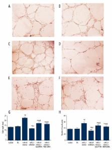
          We used an in vivo LAD rat model to simulate acute myocardial infarction. MiR-21 mimics and miR-21 inhibitors were injected and transfected into model rats in order to alter miR-21 expression. Cardiac functions were evaluated using echocardiographic measurement, ELISA, and Masson staining. In addition, lenti-PTEN and VEGF siRNA were transfected into CMEC cells using standard procedures for assessing the effect of PTEN and VEGE on cell proliferation, apoptosis, and angiogenesis. MiR-21, PTEN, and VEGF expressions were examined by RT-PCR and Western blot. The relationship between miR-21 and PTEN was determined by the luciferase activity assay.

          Results

          We demonstrated that miR-21 bonded with the 3′-UTR of PTEN and suppressed PTEN expressions. Established models significantly induced cardiac infarct volume and endothelial injury marker expressions as well as miR-21 and PTEN expressions ( P<0.05). MiR-21 mimics exhibited significantly protective effects since they down-regulated both infarction size and injury marker expressions by increasing VEGF expression and inhibiting PTEN expression ( P<0.05). In addition, results from in vitro research show that lenti-PTEN and VEGF siRNA can notably antagonize the effect of miR-21 on cell proliferation, apoptosis, and angiogenesis ( P<0.05).

          Conclusions

          MiR-21 exerts protective effects on endothelial injury through the PTEN/VEGF pathway after acute myocardial infarction.

          Related collections

          Most cited references36

          • Record: found
          • Abstract: found
          • Article: not found

          MicroRNA-92a controls angiogenesis and functional recovery of ischemic tissues in mice.

          MicroRNAs (miRs) are small noncoding RNAs that regulate gene expression by binding to target messenger RNAs (mRNAs), leading to translational repression or degradation. Here, we show that the miR-17approximately92 cluster is highly expressed in human endothelial cells and that miR-92a, a component of this cluster, controls the growth of new blood vessels (angiogenesis). Forced overexpression of miR-92a in endothelial cells blocked angiogenesis in vitro and in vivo. In mouse models of limb ischemia and myocardial infarction, systemic administration of an antagomir designed to inhibit miR-92a led to enhanced blood vessel growth and functional recovery of damaged tissue. MiR-92a appears to target mRNAs corresponding to several proangiogenic proteins, including the integrin subunit alpha5. Thus, miR-92a may serve as a valuable therapeutic target in the setting of ischemic disease.
            Bookmark
            • Record: found
            • Abstract: found
            • Article: not found

            Cytokines in atherosclerosis: pathogenic and regulatory pathways.

            Atherosclerosis is a chronic disease of the arterial wall where both innate and adaptive immunoinflammatory mechanisms are involved. Inflammation is central at all stages of atherosclerosis. It is implicated in the formation of early fatty streaks, when the endothelium is activated and expresses chemokines and adhesion molecules leading to monocyte/lymphocyte recruitment and infiltration into the subendothelium. It also acts at the onset of adverse clinical vascular events, when activated cells within the plaque secrete matrix proteases that degrade extracellular matrix proteins and weaken the fibrous cap, leading to rupture and thrombus formation. Cells involved in the atherosclerotic process secrete and are activated by soluble factors, known as cytokines. Important recent advances in the comprehension of the mechanisms of atherosclerosis provided evidence that the immunoinflammatory response in atherosclerosis is modulated by regulatory pathways, in which the two anti-inflammatory cytokines interleukin-10 and transforming growth factor-beta play a critical role. The purpose of this review is to bring together the current information concerning the role of cytokines in the development, progression, and complications of atherosclerosis. Specific emphasis is placed on the contribution of pro- and anti-inflammatory cytokines to pathogenic (innate and adaptive) and regulatory immunity in the context of atherosclerosis. Based on our current knowledge of the role of cytokines in atherosclerosis, we propose some novel therapeutic strategies to combat this disease. In addition, we discuss the potential of circulating cytokine levels as biomarkers of coronary artery disease.
              Bookmark
              • Record: found
              • Abstract: found
              • Article: not found

              Inhibition of microRNA-92a prevents endothelial dysfunction and atherosclerosis in mice.

              MicroRNAs (miRNAs) are small noncoding RNAs that regulate protein expression at post-transcriptional level. We hypothesized that a specific pool of endothelial miRNAs could be selectively regulated by flow conditions and inflammatory signals, and as such be involved in the development of atherosclerosis. To identify miRNAs, called atheromiRs, which are selectively regulated by shear stress and oxidized low-density lipoproteins (oxLDL), and to determine their role in atherogenesis. Large-scale miRNA profiling in HUVECs identified miR-92a as an atheromiR candidate, whose expression is preferentially upregulated by the combination of low shear stress (SS) and atherogenic oxLDL. Ex vivo analysis of atheroprone and atheroprotected areas of mouse arteries and human atherosclerotic plaques demonstrated the preferential expression of miR-92a in atheroprone low SS regions. In Ldlr(-/-) mice, miR-92a expression was markedly enhanced by hypercholesterolemia, in particular in atheroprone areas of the aorta. Assessment of endothelial inflammation in gain- and loss-of-function experiments targeting miR-92a expression revealed that miR-92a regulated endothelial cell activation by oxLDL, more specifically under low SS conditions, which was associated with modulation of Kruppel-like factor 2 (KLF2), Kruppel-like factor 4 (KLF4), and suppressor of cytokine signaling 5. miR-92a expression was regulated by signal transducer and activator of transcription 3 in SS- and oxLDL-dependent manner. Furthermore, specific in vivo blockade of miR-92a expression in Ldlr(-/-) mice reduced endothelial inflammation and altered the development of atherosclerosis, decreasing plaque size and promoting a more stable lesion phenotype. Upregulation of miR-92a by oxLDL in atheroprone areas promotes endothelial activation and the development of atherosclerotic lesions. Therefore, miR-92a antagomir seems as a new atheroprotective therapeutic strategy.
                Bookmark

                Author and article information

                Journal
                Med Sci Monit
                Med. Sci. Monit
                Medical Science Monitor
                Medical Science Monitor : International Medical Journal of Experimental and Clinical Research
                International Scientific Literature, Inc.
                1234-1010
                1643-3750
                2016
                06 October 2016
                : 22
                : 3562-3575
                Affiliations
                [1 ]Department of Cardiology, Xiangyang Central Hospital, Affiliated Hospital of Hubei University of Arts and Science, Xiangyang, Hubei, P.R. China
                [2 ]Department of Respiratory Medicine, Xiangyang Central Hospital, Affiliated Hospital of Hubei University of Arts and Science, Xiangyang, Hubei, P.R. China
                Author notes
                Corresponding Author: Kezhong Ma, e-mail: kezhong_ma@ 123456163.com
                [A]

                Study Design

                [B]

                Data Collection

                [C]

                Statistical Analysis

                [D]

                Data Interpretation

                [E]

                Manuscript Preparation

                [F]

                Literature Search

                [G]

                Funds Collection

                Article
                897773
                10.12659/MSM.897773
                5056537
                27708252
                7ba26309-3cb6-485b-a10c-264d2671850a
                © Med Sci Monit, 2016

                This work is licensed under a Creative Commons Attribution-NonCommercial-NoDerivs 3.0 Unported License

                History
                : 26 January 2016
                : 17 February 2016
                Categories
                Animal Study

                angiogenesis inhibitors,apoptosis,cell proliferation,myocardial infarction,pten phosphohydrolase,vascular endothelial growth factor, endocrine-gland-derived

                Comments

                Comment on this article