18
views
0
recommends
+1 Recommend
0 collections
    0
    shares
      • Record: found
      • Abstract: found
      • Article: found
      Is Open Access

      Isolation of equine herpesvirus 3 (EHV-3) from equine coital exanthema of two stallions and sero-epidemiology of EHV-3 infection in Japan

      research-article

      Read this article at

      Bookmark
          There is no author summary for this article yet. Authors can add summaries to their articles on ScienceOpen to make them more accessible to a non-specialist audience.

          Abstract

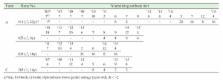
          In the spring of 2015, two stallions reared in Farms A and B in Hokkaido in Japan showed symptoms of equine coital exanthema. Equine herpesvirus 3 (EHV-3) was isolated from penis swab samples of both stallions, and the isolates from each stallion in Farms A and B were designated as SS-1 and YS-1 strains, respectively. BamHI restriction profiles of SS-1 and Japanese reference strain Iwate-1 were indistinguishable, but the BamHI-A fragment of YS-1 was larger than those of SS-1 and Iwate-1 by 1.9 kbp because of the lack of two BamHI sites. Nucleotide sequence analyses of glycoprotein G (gG), gB, gC and VP13/14 coding regions revealed that SS-1 and YS-1 had 99.77% to 100% identities to each other. These results suggested that the origins of SS-1 and YS-1 were different. For a sero-epidemiological survey, serum neutralizing tests using SS-1 against 319 sera of horses from eight farms in Hokkaido were conducted. Six of the eight farms were EHV-3 antibody-positive, and positive rates ranged from 2.6% to 17.6%. To determine the infection time of four EHV-3 antibody-positive horses, a retrospective study was conducted. Infection time of the four horses was in the breeding season, and re-infection or reactivation of latently infected EHV-3 might have occurred in one horse. However, these four horses had never shown any clinical symptoms. The results suggested that several EHV-3 strains are distributed in Japan and that infection is maintained widely in horses without clinical symptoms.

          Related collections

          Most cited references17

          • Record: found
          • Abstract: found
          • Article: not found

          Effects of bovine herpesvirus type 1 infection in calves with maternal antibodies on immune response and virus latency.

          The presence of maternally derived antibodies can interfere with the development of an active antibody response to antigen. Infection of seven passively immunized young calves with a virulent strain of bovine herpesvirus type 1 (BHV-1) was performed to determine whether they could become seronegative after the disappearance of maternal antibodies while latently infected with BHV-1. Four uninfected calves were controls. All calves were monitored serologically for 13 to 18 months. In addition, the development of a cell-mediated immune response was assessed by an in vitro antigen-specific gamma interferon (IFN-gamma) production assay. All calves had positive IFN-gamma responses as early as 7 days until at least 10 weeks after infection. However, no antibody rise was observed after infection in the three calves with the highest titers of maternal antibodies. One of the three became seronegative by virus neutralization test at 7 months of age like the control animals. This calf presented negative IFN-gamma results at the same time and was classified seronegative by enzyme-linked immunosorbent assay at around 10 months of age. This calf was latently infected, as proven by virus reexcretion after dexamethasone treatment at the end of the experiment. In conclusion, this study demonstrated that BHV-1-seronegative latent carriers can be obtained experimentally. In addition, the IFN-gamma assay was able to discriminate calves possessing only passively acquired antibodies from those latently infected by BHV-1, but it could not detect seronegative latent carriers. The failure to easily detect such animals presents an epidemiological threat for the control of BHV-1 infection.
            Bookmark
            • Record: found
            • Abstract: found
            • Article: found
            Is Open Access

            Replication characteristics of equine herpesvirus 1 and equine herpesvirus 3: comparative analysis using ex vivo tissue cultures

            Replication kinetics and invasion characteristics of equine herpesvirus-1 and -3 (EHV-1/-3) in nasal and vaginal mucosae were compared using explants. The explants were cultured during 96 h with little change in viability. The tissues were inoculated with EHV-1 03P37 (neuropathogenic), 97P70 (abortigenic) and EHV-3 04P57, collected at 0, 24, 48 and 72 h post inoculation (pi) and stained for viral antigens. Both EHV-1 and EHV-3 replicated in a plaquewise manner. The plaques were already observed at 24 h pi, their size increased over time and did not directly cross the basement membrane (BM). However, EHV-1 infected the monocytic cells (MC) and hijacked these cells to invade the lamina propria. In contrast, EHV-3 replication was fully restricted to epithelial cells; the virus did not breach the BM via a direct cell-to-cell spread nor used infected MC. EHV-1-induced plaques were larger in nasal mucosa compared to vaginal mucosa. The opposite was found for EHV-3-induced plaques. Both EHV-1 strains replicated with comparable kinetics in nasal mucosa. However, the extent of replication of the abortigenic strain in vaginal mucosa was significantly higher than that of the neuropathogenic strain. Two-to-five-fold lower numbers of EHV-1-infected MC underneath the BM were found in vaginal mucosa than in nasal mucosa. Our study has shown that (i) EHV-1 has developed in evolution a predisposition for respiratory mucosa and EHV-3 for vaginal mucosa, (ii) abortigenic EHV-1 replicates better in vaginal mucosa than neuropathogenic EHV-1 and (iii) EHV-3 demonstrated a strict epithelial tropism whereas EHV-1 in addition hijacked MC to invade the lamina propria.
              Bookmark
              • Record: found
              • Abstract: found
              • Article: not found

              Serological survey of equine viral diseases in Mongolia

              ABSTRACT Three hundred sera were collected from horses in various parts of Mongolia in 2007 and seroepidemiological surveys for several equine viruses performed on them. Equid herpesvirus 1 and equine rhinitis A virus were prevalent, and equine arteritis virus and equid herpesvirus 3 were detected over a wide area though their rates of antibody‐positivity were not high. Equine infectious anemia was distributed locally. The rates of horses antibody‐positive for Japanese encephalitis virus and equine influenza virus were low, but these were detected. Bovine coronavirus antibodies were detected at a high rate, but it was not clear whether they were due to horse coronavirus.
                Bookmark

                Author and article information

                Journal
                J Vet Med Sci
                J. Vet. Med. Sci
                JVMS
                The Journal of Veterinary Medical Science
                The Japanese Society of Veterinary Science
                0916-7250
                1347-7439
                27 January 2017
                March 2017
                : 79
                : 3
                : 636-643
                Affiliations
                [1) ]Laboratory of Veterinary Virology, Department of Pathobiology, School of Veterinary Medicine, Rakuno Gakuen University, 582 Bunkyoudai-Midorimachi, Ebetsu, Hokkaido 069-8501, Japan
                [2) ]Shadai Stallion Station, 275 Hayakita-Genbu, Abira-cho, Yufutsu-gun, Hokkaido 059-1501, Japan
                Author notes
                [* ]Correspondence to: Kirisawa, R. Laboratory of Veterinary Virology, Department of Pathobiology, School of Veterinary Medicine, Rakuno Gakuen University, 582 Bunkyoudai-Midorimachi, Ebetsu, Hokkaido 069-8501, Japan. e-mail: r-kirisa@ 123456rakuno.ac.jp
                Article
                16-0518
                10.1292/jvms.16-0518
                5383190
                28132964
                7caaba77-4dd1-47a9-a70f-4f811ae3f162
                ©2017 The Japanese Society of Veterinary Science

                This is an open-access article distributed under the terms of the Creative Commons Attribution Non-Commercial No Derivatives (by-nc-nd) License. (CC-BY-NC-ND 4.0: https://creativecommons.org/licenses/by-nc-nd/4.0/ )

                History
                : 07 October 2016
                : 11 January 2017
                Categories
                Virology
                Full Paper

                ece,ehv-3,isolation,sero-epidemiology,stallion
                ece, ehv-3, isolation, sero-epidemiology, stallion

                Comments

                Comment on this article