7
views
0
recommends
+1 Recommend
0 collections
    0
    shares
      • Record: found
      • Abstract: found
      • Article: found
      Is Open Access

      Elongated styloid process evaluation on digital panoramic radiograph in a North Italian population

      research-article

      Read this article at

      Bookmark
          There is no author summary for this article yet. Authors can add summaries to their articles on ScienceOpen to make them more accessible to a non-specialist audience.

          Abstract

          Background

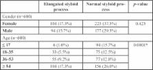
          The aim of this study is to evaluate the prevalence of elongated styloid process in digital panoramic radiographs in a North Italian population in relation to age, gender and side.

          Material and Methods

          This study was performed as a retrospective analysis on digital panoramic radiographs of 600 (271 males and 329 females) Italian patients between 6 and 87 years old. The styloid process length were measured using the measuring tool of Sidexis Software. It was measured from the point where it left the temporal bone plate to its tip. Styloid processes measuring more than 30 mm were considered elongated. Chi-squared and Fisher tests were used and the test is considered significant if the p-value is lower or equal to 0.05.

          Results

          Thirty-three per cent of the patients showed an elongated styloid process. Seventeen per cent were elongated on both right and left side, fifteen point nine per cent were elongated only in one side.

          Conclusions

          The prevalence of elongated styloid process was high and a progressive increase of the elongation prevalence was found in older groups.

          Key words:Elongated styloid process, Eagle’s syndrome, panoramic radiograph.

          Related collections

          Most cited references20

          • Record: found
          • Abstract: found
          • Article: not found

          Elongated styloid process (Eagle's syndrome): a clinical study.

          Elongated styloid process can be a source of craniofacial and cervical pain and remains a diagnostic challenge to many. The aim of the study was to determine the symptomatology and various criteria for the diagnosis of an elongated styloid process and its management. Our clinical study consisted of 58 patients with elongated styloid process who had symptoms of vague cervicofacial pain and presented to our department during a period of 10 years. Special emphasis is given to palpation of the tonsillar fossa, lidocaine infiltration test, and orthopantomography. Minimal complications and zero incidence of deep neck infection were noted. All of the patients were managed surgically through the intraoral approach, which was found to be a safe procedure. Copyright 2002 American Association of Oral and Maxillofacial Surgeons J Oral Maxillofac Surg 60:171-175, 2002
            Bookmark
            • Record: found
            • Abstract: found
            • Article: not found

            Incidence of the type and calcification patterns in patients with elongated styloid process.

            We investigated the incidence of elongated styloid process (Eagle's syndrome) using panoramic radiographs taken of 860 patients referred to our clinic. Any styloid process identified was classified according to its length, type and the pattern of calcification. Fifty-nine elongated styloid processes were identified in 32 patients (3.7%), most being bilateral; 24 patients were female and eight were male (female/male ratio: 3:1). The mean age of these patients was 43 +/- 14 years (range: 18 - 78 years). Type I (elongated) was the most frequent type on both sides (42/59); and the most frequent patterns of calcification were partially calcified on the left side (18/59) and completely calcified on the right side (16/59). Only two patients were symptomatic. A corrected differential diagnosis is important to distinguish elongated styloid process from other pathologies with partially overlapping symptoms. We would recommend that clinicians consider the possibility of Eagle's syndrome when both the clinical and radiographic evidence support this diagnosis.
              Bookmark
              • Record: found
              • Abstract: found
              • Article: found
              Is Open Access

              Evaluation of the styloid process on digital panoramic radiographs

              Background: The styloid process is an anatomical structure, whose clinical importance is not well understood. Proper clinical and radiographic evaluation can detect an elongated styloid process and calcification of the stylohyoid ligament. It has been reported that 2 – 28% of the general population show radiographic evidence of mineralization of a portion of the stylohyoid chain. The elongated styloid process may be symptomatic in many cases. Panoramic radiography is the best imaging modality to view the styloid process bilaterally. Aim: To assess the styloid process on digital panoramic radiographs. Materials and Methods: The study was conducted on 500 digital panoramic radiographs available in the archives of our department as soft copies. These radiographs were taken using a digital panoramic system. The radiographic length of the styloid process was measured on both sides using the measurement toolbars on the accompanying analysis software. For statistical analysis we used the unpaired t test, Chi-square test, and one-way ANOVA test, as necessary. Results: The average length of the left styloid was 25.41 ± 6.32 mm and that of the right styloid was 25.53 ± 6.62 mm. The length of both styloids increased with age and males had longer styloids than females. Elongated styloids were present in 19.4% of the panoramic radiographs. Langlais type I elongated styloids and a partial calcification pattern were more common than others. Conclusion: Panoramic radiography is useful for detection of an elongated styloid process and / or ossification of the stylohyoid ligament in patients with or without symptoms, and helps avoid a misdiagnosis of tonsillar pain or pain of dental, pharyngeal, or muscular origin.
                Bookmark

                Author and article information

                Journal
                J Clin Exp Dent
                J Clin Exp Dent
                Medicina Oral S.L.
                Journal of Clinical and Experimental Dentistry
                Medicina Oral S.L.
                1989-5488
                1 March 2017
                March 2017
                : 9
                : 3
                : e400-e404
                Affiliations
                [1 ]Prof, Department of Neuroscience, School of Dentistry, University of Padua, Padua, Italy
                [2 ]DDS, Department of Neuroscience, School of Dentistry, University of Padua, Padua, Italy
                [3 ]Dr, Department of Management and Engineering, University of Padova
                [4 ]Prof, Department of Orthodontics, Alma Mater Studiorum University of Bologna, Bologna, Italy
                Author notes
                Department of Neuroscience School of Dentistry University of Padua Via Giustiniani 2 – 35136 - Padua, Italy , E-mail: giobruno93@ 123456gmail.com

                Conflict of interest statement:The authors have stated explicitly that there are no competitive financial interests in connection with this article. All authors declare that all procedures followed were in accordance with the ethical standards of the responsible com-mittee on human experimentation (institutional and national) and with the Helsinki Declaration of 1964 and later ver-sions. Informed consent was obtained from all patients for being included in the study. This article does not contain any studies with human or animal subjects performed by any of the authors.

                Article
                53450
                10.4317/jced.53450
                5347289
                28298982
                7d4c26be-fa89-40b9-b4ab-150f81d5154f
                Copyright: © 2017 Medicina Oral S.L.

                This is an open-access article distributed under the terms of the Creative Commons Attribution License, which permits unrestricted use, distribution, and reproduction in any medium, provided the original work is properly cited.

                History
                : 2 December 2016
                : 25 August 2016
                Categories
                Research
                Oral Medicine and Pathology

                Comments

                Comment on this article