2
views
0
recommends
+1 Recommend
0 collections
    0
    shares
      • Record: found
      • Abstract: found
      • Article: found
      Is Open Access

      Enhanced brightness of bacterial luciferase by bioluminescence resonance energy transfer

      research-article

      Read this article at

      Bookmark
          There is no author summary for this article yet. Authors can add summaries to their articles on ScienceOpen to make them more accessible to a non-specialist audience.

          Abstract

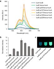
          Using the lux operon ( luxCDABE) of bacterial bioluminescence system as an autonomous luminous reporter has been demonstrated in bacteria, plant and mammalian cells. However, applications of bacterial bioluminescence-based imaging have been limited because of its low brightness. Here, we engineered the bacterial luciferase (heterodimer of luxA and luxB) by fusion with Venus, a bright variant of yellow fluorescent protein, to induce bioluminescence resonance energy transfer (BRET). By using decanal as an externally added substrate, color change and ten-times enhancement of brightness was achieved in Escherichia coli when circularly permuted Venus was fused to the C-terminus of luxB. Expression of the Venus-fused luciferase in human embryonic kidney cell lines (HEK293T) or in Nicotiana benthamiana leaves together with the substrate biosynthesis-related genes ( luxC, luxD and luxE) enhanced the autonomous bioluminescence. We believe the improved luciferase will forge the way towards the potential development of autobioluminescent reporter system allowing spatiotemporal imaging in live cells.

          Related collections

          Most cited references31

          • Record: found
          • Abstract: found
          • Article: not found

          Expanded dynamic range of fluorescent indicators for Ca(2+) by circularly permuted yellow fluorescent proteins.

          Fluorescence resonance energy transfer (FRET) technology has been used to develop genetically encoded fluorescent indicators for various cellular functions. Although most indicators have cyan- and yellow-emitting fluorescent proteins (CFP and YFP) as FRET donor and acceptor, their poor dynamic range often prevents detection of subtle but significant signals. Here, we optimized the relative orientation of the two chromophores in the Ca(2+) indicator, yellow cameleon (YC), by fusing YFP at different angles. We generated circularly permuted YFPs (cpYFPs) that showed efficient maturation and acid stability. One of the cpYFPs incorporated in YC absorbs a great amount of excited energy from CFP in its Ca(2+)-saturated form, thereby increasing the Ca(2+)-dependent change in the ratio of YFP/CFP by nearly 600%. Both in cultured cells and in the nervous system of transgenic mice, the new YC enables visualization of subcellular Ca(2+) dynamics with better spatial and temporal resolution than before. Our study provides an important guide for the development and improvement of indicators using GFP-based FRET.
            Bookmark
            • Record: found
            • Abstract: found
            • Article: not found

            A variant of yellow fluorescent protein with fast and efficient maturation for cell-biological applications.

            The green fluorescent protein (GFP) from the jellyfish Aequorea victoria has provided a myriad of applications for biological systems. Over the last several years, mutagenesis studies have improved folding properties of GFP (refs 1,2). However, slow maturation is still a big obstacle to the use of GFP variants for visualization. These problems are exacerbated when GFP variants are expressed at 37 degrees C and/or targeted to certain organelles. Thus, obtaining GFP variants that mature more efficiently is crucial for the development of expanded research applications. Among Aequorea GFP variants, yellow fluorescent proteins (YFPs) are relatively acid-sensitive, and uniquely quenched by chloride ion (Cl-). For YFP to be fully and stably fluorescent, mutations that decrease the sensitivity to both pH and Cl- are desired. Here we describe the development of an improved version of YFP named "Venus". Venus contains a novel mutation, F46L, which at 37 degrees C greatly accelerates oxidation of the chromophore, the rate-limiting step of maturation. As a result of other mutations, F64L/M153T/V163A/S175G, Venus folds well and is relatively tolerant of exposure to acidosis and Cl-. We succeeded in efficiently targeting a neuropeptide Y-Venus fusion protein to the dense-core granules of PC12 cells. Its secretion was readily monitored by measuring release of fluorescence into the medium. The use of Venus as an acceptor allowed early detection of reliable signals of fluorescence resonance energy transfer (FRET) for Ca2+ measurements in brain slices. With the improved speed and efficiency of maturation and the increased resistance to environment, Venus will enable fluorescent labelings that were not possible before.
              Bookmark
              • Record: found
              • Abstract: found
              • Article: not found

              Illuminating insights into protein-protein interactions using bioluminescence resonance energy transfer (BRET).

              Bioluminescence resonance energy transfer (BRET) is a straightforward biophysical technique for studying protein-protein interactions. It requires: (1) that proteins of interest and suitable controls be labeled with either a donor or acceptor molecule, (2) placement of these labeled proteins in the desired environment for assessing their potential interaction, and (3) use of suitable detection instrumentation to monitor resultant energy transfer. There are now several possible applications, combinations of donor and acceptor molecules, potential assay environments and detection system perturbations. Therefore, this review aims to demystify and clarify the important aspects of the BRET methodology that should be considered when using this technique.
                Bookmark

                Author and article information

                Contributors
                ng1@sanken.osaka-u.ac.jp
                Journal
                Sci Rep
                Sci Rep
                Scientific Reports
                Nature Publishing Group UK (London )
                2045-2322
                22 July 2021
                22 July 2021
                2021
                : 11
                : 14994
                Affiliations
                GRID grid.136593.b, ISNI 0000 0004 0373 3971, SANKEN (The Institute of Scientific and Industrial Research), , Osaka University, ; Ibaraki, Osaka 567-0047 Japan
                Article
                94551
                10.1038/s41598-021-94551-4
                8298465
                7da514d2-9946-4a3b-b251-9a5662415f32
                © The Author(s) 2021

                Open Access This article is licensed under a Creative Commons Attribution 4.0 International License, which permits use, sharing, adaptation, distribution and reproduction in any medium or format, as long as you give appropriate credit to the original author(s) and the source, provide a link to the Creative Commons licence, and indicate if changes were made. The images or other third party material in this article are included in the article's Creative Commons licence, unless indicated otherwise in a credit line to the material. If material is not included in the article's Creative Commons licence and your intended use is not permitted by statutory regulation or exceeds the permitted use, you will need to obtain permission directly from the copyright holder. To view a copy of this licence, visit http://creativecommons.org/licenses/by/4.0/.

                History
                : 24 February 2021
                : 6 July 2021
                Funding
                Funded by: JSPS KAKENHI
                Award ID: 17K19525
                Award Recipient :
                Categories
                Article
                Custom metadata
                © The Author(s) 2021

                Uncategorized
                molecular engineering,biochemistry,biological techniques,biophysics,biotechnology
                Uncategorized
                molecular engineering, biochemistry, biological techniques, biophysics, biotechnology

                Comments

                Comment on this article