64
views
0
recommends
+1 Recommend
0 collections
    0
    shares
      • Record: found
      • Abstract: found
      • Article: found
      Is Open Access

      Expression of oestrogen receptors, ERα, ERβ, and ERβ variants, in endometrial cancers and evidence that prostaglandin F may play a role in regulating expression of ERα

      research-article

      Read this article at

      Bookmark
          There is no author summary for this article yet. Authors can add summaries to their articles on ScienceOpen to make them more accessible to a non-specialist audience.

          Abstract

          Background

          Endometrial cancer is the most common gynaecological malignancy; risk factors include exposure to oestrogens and high body mass index. Expression of enzymes involved in biosynthesis of oestrogens and prostaglandins (PG) is often higher in endometrial cancers when compared with levels detected in normal endometrium. Oestrogens bind one of two receptors (ERα and ERβ) encoded by separate genes. The full-length receptors function as ligand-activated transcription factors; splice variant isoforms of ERβ lacking a ligand-binding domain have also been described. PGs act in an autocrine or paracrine manner by binding to specific G-protein coupled receptors.

          Methods

          We compared expression of ERs, progesterone receptor (PR) and cyclooxygenase-2 (COX-2) in stage 1 endometrial adenocarcinomas graded as well (G1), moderately (G2) or poorly (G3) differentiated (n ≥ 10 each group) using qRTPCR, single and double immunohistochemistry. We used endometrial adenocarcinoma cell lines to investigate the impact of PGF2α on expression of ERs and PR.

          Results

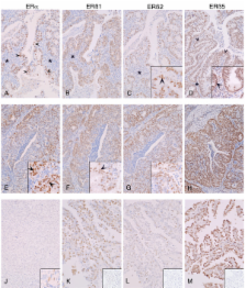
          Full length ERβ (ERβ1) and two ERβ variants (ERβ2, ERβ5) were expressed in endometrial cancers regardless of grade and the proteins were immunolocalised to the nuclei of cells in both epithelial and stromal compartments. Immunoexpression of COX-2 was most intense in cells that were ERα neg/low. Expression of PR in endometrial adenocarcinoma (Ishikawa) cell lines and tissues broadly paralleled that of ERα. Treatment of adenocarcinoma cells with PGF2α reduced expression of ERα but had no impact on ERβ1. Cells incubated with PGF2α were unable to increase expression of PR mRNA when they were incubated with E2.

          Conclusion

          We have demonstrated that ERβ5 protein is expressed in stage 1 endometrial adenocarcinomas. Expression of three ERβ variants, including the full-length protein is not grade-dependent and most cells in poorly differentiated cancers are ERβ pos/ERα neg. We found evidence of a link between COX-2, its product PGF2α, and expression of ERα and PR that sheds new light on the cross talk between steroid and PG signalling pathways in this disease.

          Related collections

          Most cited references62

          • Record: found
          • Abstract: found
          • Article: not found

          Differential ligand activation of estrogen receptors ERalpha and ERbeta at AP1 sites.

          The transactivation properties of the two estrogen receptors, ERalpha and ERbeta, were examined with different ligands in the context of an estrogen response element and an AP1 element. ERalpha and ERbeta were shown to signal in opposite ways when complexed with the natural hormone estradiol from an AP1 site: with ERalpha, 17beta-estradiol activated transcription, whereas with ERbeta, 17beta-estradiol inhibited transcription. Moreover, the antiestrogens tamoxifen, raloxifene, and Imperial Chemical Industries 164384 were potent transcriptional activators with ERbeta at an AP1 site. Thus, the two ERs signal in different ways depending on ligand and response element. This suggests that ERalpha and ERbeta may play different roles in gene regulation.
            Bookmark
            • Record: found
            • Abstract: found
            • Article: not found

            Overweight as an avoidable cause of cancer in Europe.

            There is growing evidence that excess body weight increases the risk of cancer at several sites, including kidney, endometrium, colon, prostate, gallbladder and breast in post-menopausal women. The proportion of all cancers attributable to overweight has, however, never been systematically estimated. We reviewed the epidemiological literature and quantitatively summarised, by meta-analysis, the relationship between excess weight and the risk of developing cancer at the 6 sites listed above. Estimates were then combined with sex-specific estimates of the prevalence of overweight [body mass index (BMI) 25-29 kg/m(2)] and obesity (BMI > or = 30 kg/m(2)) in each country in the European Union to obtain the proportion of cancers attributable to excess weight. Overall, excess body mass accounts for 5% of all cancers in the European Union, 3% in men and 6% in women, corresponding to 27,000 male and 45,000 female cancer cases yearly. The attributable proportion varied, in men, between 2.1% for Greece and 4.9% for Germany and, in women, between 3.9% for Denmark and 8.8% for Spain. The highest attributable proportions were obtained for cancers of the endometrium (39%), kidney (25% in both sexes) and gallbladder (25% in men and 24% in women). The largest number of attributable cases was for colon cancer (21,500 annual cases), followed by endometrium (14,000 cases) and breast (12,800 cases). Some 36,000 cases could be avoided by halving the prevalence of overweight and obese people in Europe.
              Bookmark
              • Record: found
              • Abstract: found
              • Article: not found

              Estrogen receptor (ER)-beta isoforms: a key to understanding ER-beta signaling.

              Estrogen receptor beta (ER-beta) regulates diverse physiological functions in the human body. Current studies are confined to ER-beta1, and the functional roles of isoforms 2, 4, and 5 remain unclear. Full-length ER-beta4 and -beta5 isoforms were obtained from a prostate cell line, and they exhibit differential expression in a wide variety of human tissues/cell lines. Through molecular modeling, we established that only ER-beta1 has a full-length helix 11 and a helix 12 that assumes an agonist-directed position. In ER-beta2, the shortened C terminus results in a disoriented helix 12 and marked shrinkage in the coactivator binding cleft. ER-beta4 and -beta5 completely lack helix 12. We further demonstrated that ER-beta1 is the only fully functional isoform, whereas ER-beta2, -beta4, and -beta5 do not form homodimers and have no innate activities of their own. However, the isoforms can heterodimerize with ER-beta1 and enhance its transactivation in a ligand-dependent manner. ER-beta1 tends to form heterodimers with other isoforms under the stimulation of estrogens but not phytoestrogens. Collectively, these data support the premise that (i) ER-beta1 is the obligatory partner of an ER-beta dimer, whereas the other isoforms function as variable dimer partners with enhancer activity, and (ii) a single functional helix 12 in a dimer is sufficient for gene transactivation. Thus, ER-beta behaves like a noncanonical type-I receptor, and its action may depend on differential amounts of ER-beta1 homo- and heterodimers formed upon stimulation by a specific ligand. Our findings have provided previously unrecognized directions for studying ER-beta signaling and design of ER-beta-based therapies.
                Bookmark

                Author and article information

                Journal
                BMC Cancer
                BMC Cancer
                BioMed Central
                1471-2407
                2009
                16 September 2009
                : 9
                : 330
                Affiliations
                [1 ]MRC Human Reproductive Sciences Unit, The University of Edinburgh Centre for Reproductive Biology, Queen's Medical Research Institute, 47 Little France Crescent, Edinburgh EH16 4TJ, UK
                [2 ]Division of Reproductive and Developmental Sciences, The University of Edinburgh Centre for Reproductive Biology, Queen's Medical Research Institute, 47 Little France Crescent, Edinburgh EH16 4TJ, UK
                [3 ]Division of Pathology, The University of Edinburgh, Simpson Centre for Reproductive Health, 51 Little France Crescent, Edinburgh EH16 4SA, UK
                Article
                1471-2407-9-330
                10.1186/1471-2407-9-330
                2755482
                19758455
                7da7f4ab-4682-4f5f-9b50-9b4169093456
                Copyright ©2009 Collins et al; licensee BioMed Central Ltd.

                This is an Open Access article distributed under the terms of the Creative Commons Attribution License ( http://creativecommons.org/licenses/by/2.0), which permits unrestricted use, distribution, and reproduction in any medium, provided the original work is properly cited.

                History
                : 19 May 2009
                : 16 September 2009
                Categories
                Research Article

                Oncology & Radiotherapy
                Oncology & Radiotherapy

                Comments

                Comment on this article