17
views
0
recommends
+1 Recommend
0 collections
    0
    shares
      • Record: found
      • Abstract: found
      • Article: found
      Is Open Access

      Liver Fibrosis in Non-alcoholic Fatty Liver Disease: From Liver Biopsy to Non-invasive Biomarkers in Diagnosis and Treatment

      review-article

      Read this article at

      Bookmark
          There is no author summary for this article yet. Authors can add summaries to their articles on ScienceOpen to make them more accessible to a non-specialist audience.

          Abstract

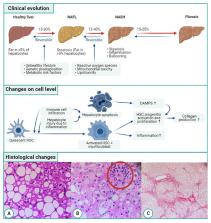
          An increasing percentage of people have or are at risk to develop non-alcoholic fatty liver disease (NAFLD) worldwide. NAFLD comprises different stadia going from isolated steatosis to non-alcoholic steatohepatitis (NASH). NASH is a chronic state of liver inflammation that leads to the transformation of hepatic stellate cells to myofibroblasts. These cells produce extra-cellular matrix that results in liver fibrosis. In a normal situation, fibrogenesis is a wound healing process that preserves tissue integrity. However, sustained and progressive fibrosis can become pathogenic. This process takes many years and is often asymptomatic. Therefore, patients usually present themselves with end-stage liver disease e.g., liver cirrhosis, decompensated liver disease or even hepatocellular carcinoma. Fibrosis has also been identified as the most important predictor of prognosis in patients with NAFLD. Currently, only a minority of patients with liver fibrosis are identified to be at risk and hence referred for treatment. This is not only because the disease is largely asymptomatic, but also due to the fact that currently liver biopsy is still the golden standard for accurate detection of liver fibrosis. However, performing a liver biopsy harbors some risks and requires resources and expertise, hence is not applicable in every clinical setting and is unsuitable for screening. Consequently, different non-invasive diagnostic tools, mainly based on analysis of blood or other specimens or based on imaging have been developed or are in development. In this review, we will first give an overview of the pathogenic mechanisms of the evolution from isolated steatosis to fibrosis. This serves as the basis for the subsequent discussion of the current and future diagnostic biomarkers and anti-fibrotic drugs.

          Related collections

          Most cited references245

          • Record: found
          • Abstract: found
          • Article: not found

          The NLRP3 inflammasome: molecular activation and regulation to therapeutics

          NLRP3 (NACHT, LRR and PYD domains-containing protein 3) is an intracellular sensor that detects a broad range of microbial motifs, endogenous danger signals and environmental irritants, resulting in the formation and activation of the NLRP3 inflammasome. Assembly of the NLRP3 inflammasome leads to caspase-1-dependent release of the proinflammatory cytokines, IL-1β and IL-18, as well as to gasdermin D-mediated pyroptotic cell death. Recent studies have revealed new regulators of the NLRP3 inflammasome, including new interacting or regulatory proteins, metabolic pathways and a regulatory mitochondrial hub. In this Review, we present the molecular, cell biological and biochemical basis of NLRP3 activation and regulation, and describe how this mechanistic understanding is leading to potential therapeutics that target the NLRP3 inflammasome.
            Bookmark
            • Record: found
            • Abstract: found
            • Article: not found

            The inflammasomes.

            Inflammasomes are molecular platforms activated upon cellular infection or stress that trigger the maturation of proinflammatory cytokines such as interleukin-1beta to engage innate immune defenses. Strong associations between dysregulated inflammasome activity and human heritable and acquired inflammatory diseases highlight the importance this pathway in tailoring immune responses. Here, we comprehensively review mechanisms directing normal inflammasome function and its dysregulation in disease. Agonists and activation mechanisms of the NLRP1, NLRP3, IPAF, and AIM2 inflammasomes are discussed. Regulatory mechanisms that potentiate or limit inflammasome activation are examined, as well as emerging links between the inflammasome and pyroptosis and autophagy. 2010 Elsevier Inc. All rights reserved.
              Bookmark
              • Record: found
              • Abstract: found
              • Article: not found

              Inflammasome-activated gasdermin D causes pyroptosis by forming membrane pores.

              Inflammatory caspases (caspases 1, 4, 5 and 11) are activated in response to microbial infection and danger signals. When activated, they cleave mouse and human gasdermin D (GSDMD) after Asp276 and Asp275, respectively, to generate an N-terminal cleavage product (GSDMD-NT) that triggers inflammatory death (pyroptosis) and release of inflammatory cytokines such as interleukin-1β. Cleavage removes the C-terminal fragment (GSDMD-CT), which is thought to fold back on GSDMD-NT to inhibit its activation. However, how GSDMD-NT causes cell death is unknown. Here we show that GSDMD-NT oligomerizes in membranes to form pores that are visible by electron microscopy. GSDMD-NT binds to phosphatidylinositol phosphates and phosphatidylserine (restricted to the cell membrane inner leaflet) and cardiolipin (present in the inner and outer leaflets of bacterial membranes). Mutation of four evolutionarily conserved basic residues blocks GSDMD-NT oligomerization, membrane binding, pore formation and pyroptosis. Because of its lipid-binding preferences, GSDMD-NT kills from within the cell, but does not harm neighbouring mammalian cells when it is released during pyroptosis. GSDMD-NT also kills cell-free bacteria in vitro and may have a direct bactericidal effect within the cytosol of host cells, but the importance of direct bacterial killing in controlling in vivo infection remains to be determined.
                Bookmark

                Author and article information

                Contributors
                Journal
                Front Med (Lausanne)
                Front Med (Lausanne)
                Front. Med.
                Frontiers in Medicine
                Frontiers Media S.A.
                2296-858X
                14 April 2021
                2021
                : 8
                : 615978
                Affiliations
                [1] 1Faculty of Health and Life Sciences, Hasselt University , Hasselt, Belgium
                [2] 2School of Nutrition and Translational Research in Metabolism, NUTRIM, Maastricht University , Maastricht, Netherlands
                [3] 3Department of Gastro-Enterology and Hepatology, Ziekenhuis Oost-Limburg , Genk, Belgium
                [4] 4Division of Gastroenterology and Hepatology, Department of Internal Medicine, Maastricht University Medical Centre , Maastricht, Netherlands
                [5] 5Department of Gastroenterology and Hepatology, University Hospital Katholieke Universiteit (KU) Leuven , Leuven, Belgium
                [6] 6Department of Gastroenterology and Hepatology, Antwerp University Hospital , Antwerp, Belgium
                [7] 7Laboratory of Experimental Medicine and Paediatrics, Faculty of Medicine and Health Sciences, University of Antwerp , Antwerp, Belgium
                Author notes

                Edited by: Peter Olinga, University of Groningen, Netherlands

                Reviewed by: Ludovico Abenavoli, University of Catanzaro, Italy; Takefumi Kimura, National Institutes of Health (NIH), United States

                *Correspondence: Sven Francque sven.francque@ 123456uza.be

                This article was submitted to Pathology, a section of the journal Frontiers in Medicine

                Article
                10.3389/fmed.2021.615978
                8079659
                33937277
                7dc89e6a-8f41-4da9-9553-bdfcb6ce0c4e
                Copyright © 2021 Heyens, Busschots, Koek, Robaeys and Francque.

                This is an open-access article distributed under the terms of the Creative Commons Attribution License (CC BY). The use, distribution or reproduction in other forums is permitted, provided the original author(s) and the copyright owner(s) are credited and that the original publication in this journal is cited, in accordance with accepted academic practice. No use, distribution or reproduction is permitted which does not comply with these terms.

                History
                : 10 October 2020
                : 22 March 2021
                Page count
                Figures: 1, Tables: 2, Equations: 0, References: 247, Pages: 20, Words: 18638
                Categories
                Medicine
                Review

                nafld,liver fibrosis,liver biopsy,non-invasive assessment,liver stiffness

                Comments

                Comment on this article