13
views
0
recommends
+1 Recommend
0 collections
    0
    shares
      • Record: found
      • Abstract: found
      • Article: found
      Is Open Access

      Skeletal intramyocellular lipid metabolism and insulin resistance

      research-article

      Read this article at

      Bookmark
          There is no author summary for this article yet. Authors can add summaries to their articles on ScienceOpen to make them more accessible to a non-specialist audience.

          Abstract

          Lipids stored in skeletal muscle cells are known as intramyocellular lipid (IMCL). Disorders involving IMCL and its causative factor, circulatory free fatty acids (FFAs), induce a toxic state and ultimately result in insulin resistance (IR) in muscle tissue. On the other hand, intramuscular triglyceride (IMTG), the most abundant component of IMCL and an essential energy source for active skeletal muscle, is different from other IMCLs, as it is stored in lipid droplets and plays a pivotal role in skeletal muscle energy homeostasis. This review discusses the association of FFA-induced ectopic lipid accumulation and IR, with specific emphasis on the relationship between IMCL/IMTG metabolism and IR.

          Related collections

          Most cited references59

          • Record: found
          • Abstract: found
          • Article: not found

          FAT SIGNALS - Lipases and Lipolysis in Lipid Metabolism and Signaling

          Lipolysis is defined as the catabolism of triacylglycerols stored in cellular lipid droplets. Recent discoveries of essential lipolytic enzymes and characterization of numerous regulatory proteins and mechanisms have fundamentally changed our perception of lipolysis and its impact on cellular metabolism. New findings that lipolytic products and intermediates participate in cellular signaling processes and that “lipolytic signaling” is particularly important in many nonadipose tissues unveil a previously underappreciated aspect of lipolysis, which may be relevant for human disease.
            Bookmark
            • Record: found
            • Abstract: found
            • Article: found
            Is Open Access

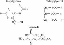
            Skeletal Muscle Triglycerides, Diacylglycerols, and Ceramides in Insulin Resistance

            OBJECTIVE Chronic exercise and obesity both increase intramyocellular triglycerides (IMTGs) despite having opposing effects on insulin sensitivity. We hypothesized that chronically exercise-trained muscle would be characterized by lower skeletal muscle diacylglycerols (DAGs) and ceramides despite higher IMTGs and would account for its higher insulin sensitivity. We also hypothesized that the expression of key skeletal muscle proteins involved in lipid droplet hydrolysis, DAG formation, and fatty-acid partitioning and oxidation would be associated with the lipotoxic phenotype. RESEARCH DESIGN AND METHODS A total of 14 normal-weight, endurance-trained athletes (NWA group) and 7 normal-weight sedentary (NWS group) and 21 obese sedentary (OBS group) volunteers were studied. Insulin sensitivity was assessed by glucose clamps. IMTGs, DAGs, ceramides, and protein expression were measured in muscle biopsies. RESULTS DAG content in the NWA group was approximately twofold higher than in the OBS group and ~50% higher than in the NWS group, corresponding to higher insulin sensitivity. While certain DAG moieties clearly were associated with better insulin sensitivity, other species were not. Ceramide content was higher in insulin-resistant obese muscle. The expression of OXPAT/perilipin-5, adipose triglyceride lipase, and stearoyl-CoA desaturase protein was higher in the NWA group, corresponding to a higher mitochondrial content, proportion of type 1 myocytes, IMTGs, DAGs, and insulin sensitivity. CONCLUSIONS Total myocellular DAGs were markedly higher in highly trained athletes, corresponding with higher insulin sensitivity, and suggest a more complex role for DAGs in insulin action. Our data also provide additional evidence in humans linking ceramides to insulin resistance. Finally, this study provides novel evidence supporting a role for specific skeletal muscle proteins involved in intramyocellular lipids, mitochondrial oxidative capacity, and insulin resistance.
              Bookmark
              • Record: found
              • Abstract: found
              • Article: not found

              Ceramide generation is sufficient to account for the inhibition of the insulin-stimulated PKB pathway in C2C12 skeletal muscle cells pretreated with palmitate.

              We have employed C2C12 myotubes to investigate lipid inhibition of insulin-stimulated signal transduction and glucose metabolism. Cells were preincubated for 18 h in the absence or presence of free fatty acids (FFAs) and stimulated with insulin, and the effects on glycogen synthesis and signaling intermediates were determined. While the unsaturated FFAs oleate and linoleate inhibited both basal and insulin-stimulated glycogen synthesis, the saturated FFA palmitate reduced only insulin-stimulated glycogen synthesis, and was found to inhibit insulin-stimulated phosphorylation of glycogen synthase kinase-3 and protein kinase B (PKB). However, no effect of palmitate was observed on tyrosine phosphorylation, p85 association, or phosphatidylinositol 3-kinase activity in IRS-1 immunoprecipitates. In contrast, palmitate promoted phosphorylation of mitogen-activated protein MAP) kinases. Ceramide, a derivative of palmitate, has recently been associated with similar inhibition of PKB, and here, ceramide levels were found to be elevated 2-fold in palmitate-treated C2C12 cells. Incubation of C2C12 cells with ceramide closely reproduced the effects of palmitate, leading to inhibition of glycogen synthesis and PKB and to stimulation of MAP kinase. We conclude that palmitate-induced insulin resistance occurs by a mechanism distinct from that of unsaturated FFAs, and involves elevation of ceramide by de novo synthesis, leading to PKB inhibition without affecting IRS-1 function.
                Bookmark

                Author and article information

                Contributors
                zhangxuelin@cupes.edu.cn
                yizc@buaa.edu.cn
                S.Cichello@latrobe.edu.au
                Journal
                Biophys Rep
                Biophys Rep
                Biophysics Reports
                Springer Berlin Heidelberg (Berlin/Heidelberg )
                2364-3439
                2364-3420
                23 October 2015
                23 October 2015
                2015
                : 1
                : 90-98
                Affiliations
                [ ]Department of Biological Science and Biotechnology, School of Biological Science and Medical Engineering, Beihang University, Beijing, 100191 China
                [ ]National Laboratory of Biomacromolecules, Institute of Biophysics, Chinese Academy of Sciences, Beijing, 100101 China
                [ ]University of Chinese Academy of Sciences, Beijing, 100049 China
                [ ]Capital University of Physical Education and Sport, Beijing, 100191 China
                [ ]School of Life Sciences, La Trobe University, Melbourne, VIC 3086 Australia
                Article
                13
                10.1007/s41048-015-0013-0
                4762133
                26942223
                7ea21b82-8694-4e1e-b60c-fa764c8e625e
                © The Author(s) 2015

                Open AccessThis article is distributed under the terms of the Creative Commons Attribution 4.0 International License ( http://creativecommons.org/licenses/by/4.0/), which permits unrestricted use, distribution, and reproduction in any medium, provided you give appropriate credit to the original author(s) and the source, provide a link to the Creative Commons license, and indicate if changes were made.

                History
                : 28 January 2015
                : 10 April 2015
                Categories
                Methods
                Custom metadata
                © The Author(s) 2015

                skeletal muscle,intramyocellular lipid,insulin resistance,free fatty acid

                Comments

                Comment on this article