2
views
0
recommends
+1 Recommend
0 collections
    0
    shares
      • Record: found
      • Abstract: found
      • Article: not found

      Restless legs syndrome secondary to pontine infarction: Clinical analysis of five cases

      research-article

      Read this article at

      ScienceOpenPublisherPMC
      Bookmark
          There is no author summary for this article yet. Authors can add summaries to their articles on ScienceOpen to make them more accessible to a non-specialist audience.

          Abstract

          Objective

          Pontine infarction is a common type of stroke in the cerebral deep structures, resulting from occlusion of small penetrating arteries, may manifest as hemi-paralysis, hemi-sensory deficit, ataxia, vertigo, and bulbar dysfunction, but patients presenting with restless legs syndrome (RLS) are extremely rare. Herein, we reported five cases with RLS as a major manifestation of pontine infarction.

          Methods

          Five cases of pontine infarction related RLS were collected from July 2013 to February 2016. The diagnosis of RLS was made according to criteria established by the International RLS Study Group (IRLSSG) in 2003. Neurological functions were assessed according to the National Institutes of Health Stroke Scale (NIHSS) and modified Rankin Scale (mRS). Severity of RLS was based on the International RLS Rating Scale (IRLS-RS). Sleep quality was assessed by Epworth Rating Scale (ERS), and individual emotional and psychological states were assessed by Hamilton Depression Scale (HDS) and Hamilton Anxiety Scale (HAS).

          Results

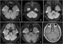
          The laboratory data at the onset including hemoglobin, serum concentration of homocysteine, blood urea nitrogen (BUN), creatinine, electrolytes, and thyroid hormones were normal. The electroencephalogram (EEG), lower-extremity somatosensory evoked potential (SEP), and nerve conduction velocity (NCV) in four limbs were normal. The average period of follow-up was 34.60 ± 12.76 months. The MRI examination showed acute or subacute pontine infarction lesions, 3 cases in the rostral inner side, 1 case in the rostral lateral and inner side, and 1 case in rostral lateral side. The neurological deficits included weakness in 4 cases, contralateral sensory deficit in 1 case, and ataxia in 2 cases. All 5 patients presented with symptom of RLS at or soon after the onset of infarction and 4 patients experienced uncomfortable sensations in the paralyzed limbs contralateral to the ischemic lesion. Their neurological deficits improved significantly 2 weeks later, but the symptoms of RLS did not resolve. Among them, 3/5 patients were treated with dopaminergic drugs. At the end of the follow-up, RLS symptom eventually resolved in 3 patients but persisted in two. The IRLS-RS, NIHSS and mRS scores were significantly lower at the onset than those at the last follow-up ( P = 0.035, 0.024 and 0.049, respectively). However, there was no significant difference in the ERS, HDS and HAS scores ( P = 0.477, 0.226 and 0.778, respectively).

          Conclusion

          RLS can be an onset manifestation of pontine infarction, clinicians should be aware of this potential symptom. RLS usually occurs in the paralyzed limbs contralateral to the infarction lesion. The pathogenesis still needs further investigation.

          Related collections

          Most cited references11

          • Record: found
          • Abstract: found
          • Article: not found

          Restless legs syndrome: diagnostic criteria, special considerations, and epidemiology. A report from the restless legs syndrome diagnosis and epidemiology workshop at the National Institutes of Health.

          Restless legs syndrome is a common yet frequently undiagnosed sensorimotor disorder. In 1995, the International Restless Legs Syndrome Study Group developed standardized criteria for the diagnosis of restless legs syndrome. Since that time, additional scientific scrutiny and clinical experience have led to a better understanding of the condition. Modification of the criteria is now necessary to better reflect that increased body of knowledge, as well as to clarify slight confusion with the wording of the original criteria. The restless legs syndrome diagnostic criteria and epidemiology workshop at the National Institutes of Health. Members of the International Restless Legs Syndrome Study Group and authorities on epidemiology and the design of questionnaires and scales. To modify the current criteria for the diagnosis of restless legs syndrome, to develop new criteria for the diagnosis of restless legs syndrome in the cognitively impaired elderly and in children, to create standardized criteria for the identification of augmentation, and to establish consistent questions for use in epidemiology studies. The essential diagnostic criteria for restless legs syndrome were developed and approved by workshop participants and the executive committee of the International Restless Legs Syndrome Study Group. Criteria were also developed and approved for the additional aforementioned groups.
            Bookmark
            • Record: found
            • Abstract: found
            • Article: not found

            Poststroke restless legs syndrome and lesion location: anatomical considerations.

            Several case studies have reported on restless legs syndrome (RLS) associated with stroke. In this study, we investigated the prevalence and the lesion topography of poststroke RLS. There were 137 patients with ischemic stroke included in this study. The diagnosis of RLS was made 1 month after the index stroke using the criteria established by the International RLS Study Group. All patients enrolled underwent magnetic resonance imaging within 7 days of the onset of the stroke. The prevalence of stroke-related RLS was calculated, and the topography of the associated ischemic lesions was analyzed. Among 137 patients, 17 patients (12.4%) were diagnosed with RLS after a stroke. Stroke-related RLS was found in 10 out of 33 patients with a basal ganglia/corona radiata infarct (30.3%), 1 out of 8 patients with an internal capsular infarct (12.5%), and 1 out of 7 patients with a thalamic infarct (14.3%). In addition, one out of 54 with a cortical lesion with/without subcortical involvement (1.9%), and 4 out of 18 patients with a pontine lesion (22.2%) had RLS. The analysis of the lesions in the cortical and subcortical group showed only 1 patient in the cortical group had stroke-related RLS, whereas 16 in the subcortical group had stroke-related RLS. The results of this study suggest that lesions of the subcortical brain areas such as the pyramidal tract and the basal ganglia-brainstem axis, which are involved in motor functions and sleep-wake cycles, may lead to RLS symptoms in patients after an ischemic stroke.
              Bookmark
              • Record: found
              • Abstract: found
              • Article: not found

              Projections of diencephalic dopamine neurons into the spinal cord in mice.

              The aim of this study is to investigate the pathway of diencephalic dopaminergic (DA) neuronal innervating into the spinal cord in mice, the pathway is postulated relevant to clinical restless legs syndrome (RLS). Tyrosine hydroxylase (TH) immunohistochemistry was used to identify the DA neuron. The fluorescent tracer Fluoro-Gold (FG) was stereotaxically injected into the T10-L5 spinal cord of CBL57 mice (n=20) seven days before the animals were sacrificed. The diencephalic sections were stained with TH antibody and the FG tracer present in the diencephalic DA neurons were examined under fluoresce microscope. The average number of total DA neurons per side in A11, A12, A13 and A14 was 66+/-8, 221+/-12, 350+/-17 and 254+/-21 respectively. After being injected into the spinal cord, FG reached the DA neurons within the A10 and A11 groups, but didn't target to any other DA neuron groups including the A8 and A9 groups in substantia nigra (SN). The diencephalic A11 DA neurons possessed long axons extending over several segments and possibly traversing the entire length of the spinal cord. It is the first time to report A10 and A11 DA neuron projections into the spinal cord in mice.
                Bookmark

                Author and article information

                Contributors
                Journal
                Chronic Dis Transl Med
                Chronic Dis Transl Med
                Chronic Diseases and Translational Medicine
                KeAi Publishing
                2095-882X
                2589-0514
                01 September 2017
                September 2017
                01 September 2017
                : 3
                : 3
                : 186-190
                Affiliations
                [a ]Department of Neurology, Capital Medical University Affiliated Beijing Friendship Hospital, Beijing 100050, China
                [b ]Department of Neurology, The Fourth Central Hospital of Tianjin, Tianjin 300140, China
                [c ]Department of Neurology, Beijing Ditan Hospital, Capital Medical University, Beijing 100015, China
                [d ]The Center for Translational Research on Neurological Diseases, The First Affiliated Hospital of Dalian Medical University, Dalian, Liaoning 116011, China
                [e ]Department of Neurology, Methodist Neurological Institute, Houston, TX 77030, USA
                Author notes
                []Corresponding author. tuohzh@ 123456sina.cn
                [∗∗ ]Corresponding author. wondo@ 123456houstonmethodist.org
                Article
                S2095-882X(17)30060-9
                10.1016/j.cdtm.2017.08.001
                5643777
                7f664e7a-7759-4b1d-8281-b0669b068796
                © 2017 Chinese Medical Association. Production and hosting by Elsevier B.V. on behalf of KeAi Communications Co., Ltd.

                This is an open access article under the CC BY-NC-ND license (http://creativecommons.org/licenses/by-nc-nd/4.0/).

                History
                : 4 July 2017
                Categories
                Original Article

                restless legs syndrome,pontine infarction,clinical features

                Comments

                Comment on this article