7
views
0
recommends
+1 Recommend
0 collections
    0
    shares
      • Record: found
      • Abstract: found
      • Article: found
      Is Open Access

      A method package for electrophysiological evaluation of reconstructed or regenerated facial nerves in rodents

      research-article

      Read this article at

      Bookmark
          There is no author summary for this article yet. Authors can add summaries to their articles on ScienceOpen to make them more accessible to a non-specialist audience.

          Graphical abstract

          Abstract

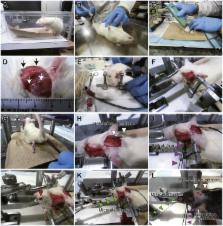
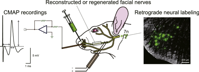
          Compound muscle action potential (CMAP) recording via reconstructed or regenerated motor axons is a critical examination to evaluate newly developed surgical and regeneration techniques. However, there is currently no documentation on technical aspects of CMAP recordings via reconstructed or regenerated facial nerves. We have studied new techniques of plastic surgery and nerve regeneration using a rat facial nerve defect model for years, standardizing an evaluation pipeline using CMAP recordings. Here we describe our CMAP recording procedure in detail as a package including surgical preparation, data acquisition, analysis and troubleshooting. Each resource is available in public repositories and is maintained as a version control system. In addition, we demonstrate that our analytical pipeline can not only be applied to rats, but also mice. Finally, we show that CMAP recordings can be practically combined with other behavioral and anatomical examinations. For example, retrograde motor neuron labeling provides anatomical evidence for physical routes between the facial motor nucleus and its periphery through reconstructed or regenerated facial nerves, in addition to electrophysiological evidence by CMAP recordings from the same animal.

          • Standardized surgical, recording and analytical procedures for the functional evaluation of reconstructed or regenerated facial nerves of rats, extended to mice.

          • The functional evaluation can be combined with anatomical evaluations.

          • The methods described here are maintained in public repositories as version control systems.

          Related collections

          Most cited references17

          • Record: found
          • Abstract: found
          • Article: not found

          Spatiotemporal dynamics of cortical sensorimotor integration in behaving mice.

          Tactile information is actively acquired and processed in the brain through concerted interactions between movement and sensation. Somatosensory input is often the result of self-generated movement during the active touch of objects, and conversely, sensory information is used to refine motor control. There must therefore be important interactions between sensory and motor pathways, which we chose to investigate in the mouse whisker sensorimotor system. Voltage-sensitive dye was applied to the neocortex of mice to directly image the membrane potential dynamics of sensorimotor cortex with subcolumnar spatial resolution and millisecond temporal precision. Single brief whisker deflections evoked highly distributed depolarizing cortical sensory responses, which began in the primary somatosensory barrel cortex and subsequently excited the whisker motor cortex. The spread of sensory information to motor cortex was dynamically regulated by behavior and correlated with the generation of sensory-evoked whisker movement. Sensory processing in motor cortex may therefore contribute significantly to active tactile sensory perception.
            Bookmark
            • Record: found
            • Abstract: found
            • Article: not found

            The musculature of the mystacial vibrissae of the white mouse.

            J Dörfl (1982)
            Striated muscles of the mystacial region of the common albino mouse have been described. They were divided into two categories: extrinsic and intrinsic. The four extrinsic muscles (m. levator labii superioris, m. maxillolabialis, m. transversus nasi, m. nasalis) belong to the facial muscles. They originate on the skull and insert into the corium between the mystacial vibrissae. Their contraction moves the whole mystacial region in directions dependent on their origins. Intrinsic (follicular) muscles are associated solely with the vibrissal follicles and have no bony attachment. They were found around follicles alpha, beta, gamma, delta, around all follicles of rows A and B, and around the first six follicles of rows C, D and E. The form of the follicular muscle is a sling connecting two adjacent follicles of the same row. The arc of the sling surrounds the inferior part of the rostral follicle and the two extremities insert to the conical body of the caudal follicle and to the neighbouring corium. They are the protractors of the vibrissae. The inferior parts of the vibrissal follicles of a given row are fixed in a fibrous band which inserts in the anterior part of the muzzle. It is proposed that these bands become stretched during the protraction of vibrissae and contract, by their elasticity, immediately upon the end of the follicular muscles' contraction, executing the fast return of vibrissae to their resting, retracted position.
              Bookmark
              • Record: found
              • Abstract: found
              • Article: not found

              Electrophysiologic and functional evaluations of regenerated facial nerve defects with a tube containing dental pulp cells in rats.

              Dental pulp tissue contains Schwann and neural progenitor cells. Tissue-engineered nerve conduits with dental pulp cells promote facial nerve regeneration in rats. However, no nerve functional or electrophysiologic evaluations were performed. This study investigated the compound muscle action potential recordings and facial functional analysis of dental pulp cell regenerated nerve in rats.
                Bookmark

                Author and article information

                Contributors
                Journal
                MethodsX
                MethodsX
                MethodsX
                Elsevier
                2215-0161
                30 March 2018
                2018
                30 March 2018
                : 5
                : 283-298
                Affiliations
                [a ]Department of Physiology I (Neurophysiology), Tokyo Women’s Medical University, Tokyo, Japan
                [b ]Department of Plastic and Reconstructive Surgery, Tokyo Women’s Medical University, Tokyo, Japan
                [c ]Department of Oral and Maxillofacial Surgery, Tokyo Women’s Medical University, Tokyo, Japan
                Author notes
                [* ]Corresponding author. Present address: MTA-SZTE “Momentum” Oscillatory Neuronal Networks Research Group, Department of Physiology, University of Szeged, Szeged 6720, Hungary. yuichi-takeuchi@ 123456umin.net
                [1]

                Department of Physiology I (Neurophysiology), School of Medicine, Tokyo Women’s Medical University, 8-1 Kawada-cho, Shinjuku-ku, Tokyo 162-8666, Japan.

                Article
                S2215-0161(18)30045-1
                10.1016/j.mex.2018.03.007
                6055010
                30042925
                838eac87-103f-433e-9163-4f8efa41955b
                © 2018 The Author(s)

                This is an open access article under the CC BY license (http://creativecommons.org/licenses/by/4.0/).

                History
                : 17 October 2017
                : 28 March 2018
                Categories
                Medicine and Dentistry

                compound muscle action potential recordings via reconstructed or regenerated facial nerves,facial nerve,reconstruction,regeneration,compound muscle action potential,retrograde tracer,rat,mouse,analysis,igor pro,matlab,version control system

                Comments

                Comment on this article