2
views
0
recommends
+1 Recommend
0 collections
    0
    shares
      • Record: found
      • Abstract: found
      • Article: found
      Is Open Access

      Assessment of a Non Invasive Brain Oximeter in Volunteers Undergoing Acute Hypoxia

      research-article
      1 , 2
      Medical Devices (Auckland, N.Z.)
      Dove
      oximetry, brain injury, brain, volunteer, hypoxia, monitor

      Read this article at

      Bookmark
          There is no author summary for this article yet. Authors can add summaries to their articles on ScienceOpen to make them more accessible to a non-specialist audience.

          Abstract

          Introduction

          Research in traumatic brain injury suggests better patient outcomes when invasive oxygen monitoring is used to detect and correct episodes of brain hypoxia. Invasive brain oxygen monitoring is, however, not routinely used due to the risks, costs and technical challengers. We are developing a non-invasive brain oximeter to address these limitations. The monitor uses the principles of pulse oximetry to record a brain photoplethysmographic waveform and oxygen saturations. We undertook a study in volunteers to assess the new monitor.

          Patients and Methods

          We compared the temporal changes in the brain and skin oxygen saturations in six volunteers undergoing progressive hypoxia to reach arterial saturations of 70%. This approach provides a method to discriminate potential contamination of the brain signal by skin oxygen levels, as the responses in brain and skin oxygen saturations are distinct due to the auto-regulation of cerebral blood flow to compensate for hypoxia. Conventional pulse oximetry was used to assess skin oxygen levels. Blood was also collected from the internal jugular vein and correlated with the brain oximeter oxygen levels.

          Results

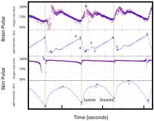
          At baseline, a photoplethysmographic waveform consistent with that expected from the brain was obtained in five subjects. The signal was adequate to assess oxygen saturations in three subjects. During hypoxia, the brain's oximeter oxygen saturation fell to 74%, while skin saturation fell to 50% (P<0.0001). The brain photoplethysmographic waveform developed a high-frequency oscillation of ~7 Hz, which was not present in the skin during hypoxia. A weak correlation between the brain oximeter and proximal internal jugular vein oxygen levels was demonstrated, R 2=0.24, P=0.01.

          Conclusion

          Brain oximeter oxygen saturations were relatively well preserved compared to the skin during hypoxia. These findings are consistent with the expected physiological responses and suggest skin oxygen levels did not markedly contaminate the brain oximeter signal.

          Related collections

          Most cited references45

          • Record: found
          • Abstract: found
          • Article: found
          Is Open Access

          The pulsating brain: A review of experimental and clinical studies of intracranial pulsatility

          The maintenance of adequate blood flow to the brain is critical for normal brain function; cerebral blood flow, its regulation and the effect of alteration in this flow with disease have been studied extensively and are very well understood. This flow is not steady, however; the systolic increase in blood pressure over the cardiac cycle causes regular variations in blood flow into and throughout the brain that are synchronous with the heart beat. Because the brain is contained within the fixed skull, these pulsations in flow and pressure are in turn transferred into brain tissue and all of the fluids contained therein including cerebrospinal fluid. While intracranial pulsatility has not been a primary focus of the clinical community, considerable data have accrued over the last sixty years and new applications are emerging to this day. Investigators have found it a useful marker in certain diseases, particularly in hydrocephalus and traumatic brain injury where large changes in intracranial pressure and in the biomechanical properties of the brain can lead to significant changes in pressure and flow pulsatility. In this work, we review the history of intracranial pulsatility beginning with its discovery and early characterization, consider the specific technologies such as transcranial Doppler and phase contrast MRI used to assess various aspects of brain pulsations, and examine the experimental and clinical studies which have used pulsatility to better understand brain function in health and with disease.
            Bookmark
            • Record: found
            • Abstract: found
            • Article: not found

            Brain Oxygen Optimization in Severe Traumatic Brain Injury Phase-II

            A relationship between reduced brain tissue oxygenation and poor outcome following severe traumatic brain injury has been reported in observational studies. We designed a Phase II trial to assess whether a neurocritical care management protocol could improve brain tissue oxygenation levels in patients with severe traumatic brain injury and the feasibility of a Phase III efficacy study.
              Bookmark
              • Record: found
              • Abstract: found
              • Article: not found

              Reduced mortality rate in patients with severe traumatic brain injury treated with brain tissue oxygen monitoring.

              An intracranial pressure (ICP) monitor, from which cerebral perfusion pressure (CPP) is estimated, is recommended in the care of severe traumatic brain injury (TBI). Nevertheless, optimal ICP and CPP management may not always prevent cerebral ischemia, which adversely influences patient outcome. The authors therefore determined whether the addition of a brain tissue oxygen tension (PO2) monitor in the treatment of TBI was associated with an improved patient outcome. Patients with severe TBI (Glasgow Coma Scale [GCS] score < 8) who had been admitted to a Level I trauma center were evaluated as part of a prospective observational database. Patients treated with ICP and brain tissue PO2 monitoring were compared with historical controls matched for age, pathological features, admission GCS score, and Injury Severity Score who had undergone ICP monitoring alone. Therapy in both patient groups was aimed at maintaining an ICP less than 20 mm Hg and a CPP greater than 60 mm Hg. Among patients whose brain tissue PO2 was monitored, oxygenation was maintained at levels greater than 25 mm Hg. Twenty-five patients with a mean age of 44 +/- 14 years were treated using an ICP monitor alone. Twenty-eight patients with a mean age of 38 +/- 18 years underwent brain tissue PO2-directed care. The mean daily ICP and CPP levels were similar in each group. The mortality rate in patients treated using conventional ICP and CPP management was 44%. Patients who also underwent brain tissue PO2 monitoring had a significantly reduced mortality rate of 25% (p < 0.05). The use of both ICP and brain tissue PO2 monitors and therapy directed at brain tissue PO2 is associated with reduced patient death following severe TBI.
                Bookmark

                Author and article information

                Journal
                Med Devices (Auckl)
                Med Devices (Auckl)
                MDER
                mder
                Medical Devices (Auckland, N.Z.)
                Dove
                1179-1470
                30 June 2020
                2020
                : 13
                : 183-194
                Affiliations
                [1 ]Cyban Pty Ltd , Melbourne, VIC, Australia
                [2 ]Human Pharmacology and Physiology Laboratory, Department of Anesthesiology and School of Nursing, Duke University , Durham, NC, USA
                Author notes
                Correspondence: Barry Dixon Tel +61 0439618815 Email barry.dixon@cyban.com.au
                Author information
                http://orcid.org/0000-0002-5151-1674
                http://orcid.org/0000-0002-5989-3961
                Article
                250102
                10.2147/MDER.S250102
                7335769
                8425273f-141f-467b-9fc4-6d9d5ace139d
                © 2020 Dixon and MacLeod.

                This work is published and licensed by Dove Medical Press Limited. The full terms of this license are available at https://www.dovepress.com/terms.php and incorporate the Creative Commons Attribution – Non Commercial (unported, v3.0) License ( http://creativecommons.org/licenses/by-nc/3.0/). By accessing the work you hereby accept the Terms. Non-commercial uses of the work are permitted without any further permission from Dove Medical Press Limited, provided the work is properly attributed. For permission for commercial use of this work, please see paragraphs 4.2 and 5 of our Terms ( https://www.dovepress.com/terms.php).

                History
                : 16 February 2020
                : 09 June 2020
                Page count
                Figures: 8, Tables: 1, References: 48, Pages: 12
                Funding
                Funded by: Cyban Pty Ltd
                This research received no specific grant from any funding agency in the public or not-for-profit sectors and was funded by Cyban Pty Ltd.
                Categories
                Original Research

                Biotechnology
                oximetry,brain injury,brain,volunteer,hypoxia,monitor
                Biotechnology
                oximetry, brain injury, brain, volunteer, hypoxia, monitor

                Comments

                Comment on this article