8
views
0
recommends
+1 Recommend
0 collections
    0
    shares
      • Record: found
      • Abstract: found
      • Article: found
      Is Open Access

      Structural investigation of a chaperonin in action reveals how nucleotide binding regulates the functional cycle

      research-article

      Read this article at

      Bookmark
          There is no author summary for this article yet. Authors can add summaries to their articles on ScienceOpen to make them more accessible to a non-specialist audience.

          Abstract

          Site-selective isotope labeling enables structural and functional investigation of a working 1-MDa chaperonin by NMR spectroscopy.

          Abstract

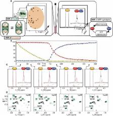
          Chaperonins are ubiquitous protein assemblies present in bacteria, eukaryota, and archaea, facilitating the folding of proteins, preventing protein aggregation, and thus participating in maintaining protein homeostasis in the cell. During their functional cycle, they bind unfolded client proteins inside their double ring structure and promote protein folding by closing the ring chamber in an adenosine 5′-triphosphate (ATP)–dependent manner. Although the static structures of fully open and closed forms of chaperonins were solved by x-ray crystallography or electron microscopy, elucidating the mechanisms of such ATP-driven molecular events requires studying the proteins at the structural level under working conditions. We introduce an approach that combines site-specific nuclear magnetic resonance observation of very large proteins, enabled by advanced isotope labeling methods, with an in situ ATP regeneration system. Using this method, we provide functional insight into the 1-MDa large hsp60 chaperonin while processing client proteins and reveal how nucleotide binding, hydrolysis, and release control switching between closed and open states. While the open conformation stabilizes the unfolded state of client proteins, the internalization of the client protein inside the chaperonin cavity speeds up its functional cycle. This approach opens new perspectives to study structures and mechanisms of various ATP-driven biological machineries in the heat of action.

          Related collections

          Most cited references45

          • Record: found
          • Abstract: found
          • Article: not found

          Heat Shock Proteins and Cancer.

          Heat shock proteins (HSPs) constitute a large family of proteins involved in protein folding and maturation whose expression is induced by heat shock or other stressors. The major groups are classified based on their molecular weights and include HSP27, HSP40, HSP60, HSP70, HSP90, and large HSPs. HSPs play a significant role in cellular proliferation, differentiation, and carcinogenesis. In this article we comprehensively review the roles of major HSPs in cancer biology and pharmacology. HSPs are thought to play significant roles in the molecular mechanisms leading to cancer development and metastasis. HSPs may also have potential clinical uses as biomarkers for cancer diagnosis, for assessing disease progression, or as therapeutic targets for cancer therapy.
            Bookmark
            • Record: found
            • Abstract: found
            • Article: not found

            Cross-correlated relaxation enhanced 1H[bond]13C NMR spectroscopy of methyl groups in very high molecular weight proteins and protein complexes.

            A comparison of HSQC and HMQC pulse schemes for recording (1)H[bond](13)C correlation maps of protonated methyl groups in highly deuterated proteins is presented. It is shown that HMQC correlation maps can be as much as a factor of 3 more sensitive than their HSQC counterparts and that the sensitivity gains result from a TROSY effect that involves cancellation of intra-methyl dipolar relaxation interactions. (1)H[bond](13)C correlation spectra are recorded on U-[(15)N,(2)H], Ile delta 1-[(13)C,(1)H] samples of (i) malate synthase G, a 723 residue protein, at 37 and 5 degrees C, and of (ii) the protease ClpP, comprising 14 identical subunits, each with 193 residues (305 kDa), at 5 degrees C. The high quality of HMQC spectra obtained in short measuring times strongly suggests that methyl groups will be useful probes of structure and dynamics in supramolecular complexes.
              Bookmark
              • Record: found
              • Abstract: found
              • Article: not found

              Quantitative dynamics and binding studies of the 20S proteasome by NMR.

              The machinery used by the cell to perform essential biological processes is made up of large molecular assemblies. One such complex, the proteasome, is the central molecular machine for removal of damaged and misfolded proteins from the cell. Here we show that for the 670-kilodalton 20S proteasome core particle it is possible to overcome the molecular weight limitations that have traditionally hampered quantitative nuclear magnetic resonance (NMR) spectroscopy studies of such large systems. This is achieved by using an isotope labelling scheme where isoleucine, leucine and valine methyls are protonated in an otherwise highly deuterated background in concert with experiments that preserve the lifetimes of the resulting NMR signals. The methodology has been applied to the 20S core particle to reveal functionally important motions and interactions by recording spectra on complexes with molecular weights of up to a megadalton. Our results establish that NMR spectroscopy can provide detailed insight into supra-molecular structures over an order of magnitude larger than those routinely studied using methodology that is generally applicable.
                Bookmark

                Author and article information

                Journal
                Sci Adv
                Sci Adv
                SciAdv
                advances
                Science Advances
                American Association for the Advancement of Science
                2375-2548
                September 2018
                19 September 2018
                : 4
                : 9
                : eaau4196
                Affiliations
                University of Grenoble Alpes, CNRS, CEA, IBS, F-38000 Grenoble, France.
                Author notes
                [*]

                Present address: Biozentrum University of Basel, Klingelbergstrasse 70, 4056 Basel, Switzerland.

                [†]

                Present address: NMR-Bio, F-38000 Grenoble, France.

                []Corresponding author. Email: macek@ 123456nmr-bio.com (P.M.); paul.schanda@ 123456ibs.fr (P.S.); jerome.boisbouvier@ 123456ibs.fr (J.B.)
                Author information
                http://orcid.org/0000-0002-2351-0455
                http://orcid.org/0000-0003-1510-2846
                http://orcid.org/0000-0002-8279-3053
                http://orcid.org/0000-0002-1459-3201
                http://orcid.org/0000-0003-3774-7717
                http://orcid.org/0000-0002-9350-7606
                http://orcid.org/0000-0003-3278-3639
                Article
                aau4196
                10.1126/sciadv.aau4196
                6154984
                30255156
                84c6c86d-a5e8-48a2-9e02-99f0ad6d57d6
                Copyright © 2018 The Authors, some rights reserved; exclusive licensee American Association for the Advancement of Science. No claim to original U.S. Government Works. Distributed under a Creative Commons Attribution NonCommercial License 4.0 (CC BY-NC).

                This is an open-access article distributed under the terms of the Creative Commons Attribution-NonCommercial license, which permits use, distribution, and reproduction in any medium, so long as the resultant use is not for commercial advantage and provided the original work is properly cited.

                History
                : 08 June 2018
                : 01 August 2018
                Funding
                Funded by: doi http://dx.doi.org/10.13039/501100000781, European Research Council;
                Award ID: ERC-CoG-2010-260887
                Funded by: doi http://dx.doi.org/10.13039/501100000781, European Research Council;
                Award ID: ERC-StG-2012-311318
                Funded by: doi http://dx.doi.org/10.13039/501100001665, Agence Nationale de la Recherche;
                Award ID: ANR-10-INSB-05-02
                Funded by: doi http://dx.doi.org/10.13039/501100001665, Agence Nationale de la Recherche;
                Award ID: ANR-10-LABX-49-01
                Funded by: Fondation Recherche Medical;
                Funded by: Fonds FEDER;
                Funded by: GIS-Infrastrutures en Biologie Santé et Agronomie;
                Categories
                Research Article
                Research Articles
                SciAdv r-articles
                Biophysics
                Structural Biology
                Structural Biology
                Custom metadata
                Eunice Ann Alesin

                Comments

                Comment on this article