11
views
0
recommends
+1 Recommend
0 collections
    0
    shares
      • Record: found
      • Abstract: found
      • Article: found
      Is Open Access

      Plasma DNA Mediate Autonomic Dysfunctions and White Matter Injuries in Patients with Parkinson's Disease

      research-article

      Read this article at

      Bookmark
          There is no author summary for this article yet. Authors can add summaries to their articles on ScienceOpen to make them more accessible to a non-specialist audience.

          Abstract

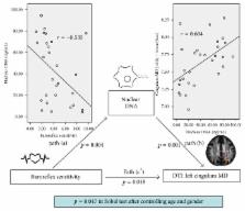
          Background. Cardiovascular autonomic dysfunction is well known in Parkinson's disease (PD) presentation and it produces hypoperfusion of vital organs. The association between cardiovascular autonomic dysfunction and oxidative stress was examined in previous animal models. Oxidative stress and neuroinflammation were thought to have roles in PD pathogenesis. Owing to the relative low intrinsic antioxidative properties, brain white matter (WM) is vulnerable to the oxidative stress. This study is conducted to examine possible relationships by using a hypothesis-driven mediation model. Methods. Twenty-nine patients with PD and 26 healthy controls participated in this study, with complete examinations of cardiac autonomic parameters, plasma DNA level, and WM integrity. A single-level three-variable mediation model was used to investigate the possible relationships. Results. The elevated serum oxidative stress biomarkers include plasma nuclear DNA and mitochondrial DNA, and poorer cardiac autonomic parameters and multiple regional microstructural WM changes are demonstrated. Further mediation analysis shows that plasma nuclear DNA served as the mediators between poorer baroreflex sensitivity and mean diffusivity changes in cingulum. Conclusions. These results provide a possible pathophysiology for how the poor baroreflex sensitivity and higher oxidative stress adversely impacted the WM integrity. This model could provide us with a piece of the puzzle of the entire PD pathogenesis.

          Related collections

          Most cited references38

          • Record: found
          • Abstract: found
          • Article: not found

          Demyelination increases radial diffusivity in corpus callosum of mouse brain.

          Myelin damage, as seen in multiple sclerosis (MS) and other demyelinating diseases, impairs axonal conduction and can also be associated with axonal degeneration. Accurate assessments of these conditions may be highly beneficial in evaluating and selecting therapeutic strategies for patient management. Recently, an analytical approach examining diffusion tensor imaging (DTI) derived parameters has been proposed to assess the extent of axonal damage, demyelination, or both. The current study uses the well-characterized cuprizone model of experimental demyelination and remyelination of corpus callosum in mouse brain to evaluate the ability of DTI parameters to detect the progression of myelin degeneration and regeneration. Our results demonstrate that the extent of increased radial diffusivity reflects the severity of demyelination in corpus callosum of mouse brain affected by cuprizone treatment. Subsequently, radial diffusivity decreases with the progression of remyelination. Furthermore, radial diffusivity changes were specific to the time course of changes in myelin integrity as distinct from axonal injury, which was detected by betaAPP immunostaining and shown to be most extensive prior to demyelination. Radial diffusivity offers a specific assessment of demyelination and remyelination, as distinct from acute axonal damage.
            Bookmark
            • Record: found
            • Abstract: found
            • Article: not found

            Principles of diffusion tensor imaging and its applications to basic neuroscience research.

            The brain contains more than 100 billion neurons that communicate with each other via axons for the formation of complex neural networks. The structural mapping of such networks during health and disease states is essential for understanding brain function. However, our understanding of brain structural connectivity is surprisingly limited, due in part to the lack of noninvasive methodologies to study axonal anatomy. Diffusion tensor imaging (DTI) is a recently developed MRI technique that can measure macroscopic axonal organization in nervous system tissues. In this article, the principles of DTI methodologies are explained, and several applications introduced, including visualization of axonal tracts in myelin and axonal injuries as well as human brain and mouse embryonic development. The strengths and limitations of DTI and key areas for future research and development are also discussed.
              Bookmark
              • Record: found
              • Abstract: found
              • Article: not found

              Diffusion tensor imaging of cingulum fibers in mild cognitive impairment and Alzheimer disease.

              Neuroimaging in mild cognitive impairment (MCI) and Alzheimer disease (AD) generally shows medial temporal lobe atrophy and diminished glucose metabolism and cerebral blood flow in the posterior cingulate gyrus. However, it is unclear whether these abnormalities also impact the cingulum fibers, which connect the medial temporal lobe and the posterior cingulate regions. To use diffusion tensor imaging (DTI), by measuring fractional anisotropy (FA), to test 1) if MCI and AD are associated with DTI abnormalities in the parahippocampal and posterior cingulate regions of the cingulum fibers; 2) if white matter abnormalities extend to the neocortical fiber connections in the corpus callosum (CC); 3) if DTI improves accuracy to separate AD and MCI from healthy aging vs structural MRI. DTI and structural MRI were preformed on 17 patients with AD, 17 with MCI, and 18 cognitively normal (CN) subjects. FA of the cingulum fibers was significantly reduced in MCI, and even more in AD. FA was also significantly reduced in the splenium of the CC in AD, but not in MCI. Adding DTI to hippocampal volume significantly improved the accuracy to separate MCI and AD from CN. Assessment of the cingulum fibers using diffusion tensor imaging may aid early diagnosis of Alzheimer disease.
                Bookmark

                Author and article information

                Journal
                Oxid Med Cell Longev
                Oxid Med Cell Longev
                OMCL
                Oxidative Medicine and Cellular Longevity
                Hindawi Publishing Corporation
                1942-0900
                1942-0994
                2017
                23 January 2017
                : 2017
                : 7371403
                Affiliations
                1Department of Diagnostic Radiology, Kaohsiung Chang Gung Memorial Hospital, Kaohsiung, Taiwan
                2Chang Gung University College of Medicine, Kaohsiung, Taiwan
                3Department of Neurology, Kaohsiung Chang Gung Memorial Hospital, Kaohsiung, Taiwan
                4Graduate Institute of Medical Mechatronics, Chang Gung University, Taoyuan, Taiwan
                5Department of Neurology, Chang Gung Memorial Hospital, Linkou, Taoyuan, Taiwan
                6Department of Medicine, Kaohsiung Chang Gung Memorial Hospital, Kaohsiung, Taiwan
                Author notes

                Academic Editor: James Duce

                Author information
                http://orcid.org/0000-0003-0201-9015
                http://orcid.org/0000-0002-5716-3930
                http://orcid.org/0000-0002-7140-9132
                http://orcid.org/0000-0002-5258-8569
                Article
                10.1155/2017/7371403
                5292395
                28232858
                862184f2-7eb7-4537-9c91-efac1e6c3a8d
                Copyright © 2017 Meng-Hsiang Chen et al.

                This is an open access article distributed under the Creative Commons Attribution License, which permits unrestricted use, distribution, and reproduction in any medium, provided the original work is properly cited.

                History
                : 1 September 2016
                : 8 November 2016
                : 19 December 2016
                Funding
                Funded by: National Science Council
                Award ID: MOST 103-2314-B-182A-010-MY3
                Award ID: MOST 104-2314-B-182A-053
                Funded by: Chang Gung Memorial Hospital
                Award ID: CMRPG891511
                Award ID: CMRPG8B0831
                Award ID: CMRPG890801
                Award ID: CMRPG8C0021
                Award ID: CMRPG8E0621
                Categories
                Research Article

                Molecular medicine
                Molecular medicine

                Comments

                Comment on this article