3
views
0
recommends
+1 Recommend
0 collections
    0
    shares
      • Record: found
      • Abstract: found
      • Article: found
      Is Open Access

      3D-Visualization of Neurovascular Compression at the Ventrolateral Medulla in Patients with Arterial Hypertension

      research-article

      Read this article at

      Bookmark
          There is no author summary for this article yet. Authors can add summaries to their articles on ScienceOpen to make them more accessible to a non-specialist audience.

          Abstract

          Purpose

          Controversy exists on the association of arterial hypertension (HTN) and neurovascular compression (NVC) at the ventrolateral medulla (VLM). No standardized and reproducible technique has been introduced yet for detection of NVC in HTN. This study aimed to generate, analyze and compare different results of exact reproducible anatomical 3D-representations of the VLM in patients with HTN, based on magnetic resonance imaging (MRI).

          Methods

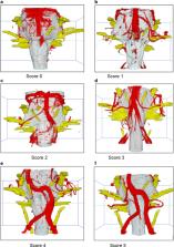
          A 3T scanner provided MRI (T2-constructive interference in steady state (CISS) high resolution imaging and three-dimensional Time-of-flight (3D-TOF) angiography) from the posterior fossa of 44 patients with clinical treatment-resistant HTN. Image processing consists of segmentation of the CISS data, registration and fusion of the CISS and TOF data and visualization. For each patient two 3D-visualizations (before and after fusion) were obtained. The reproduction quality of the vessels, flow-related signal variability and pulsation artifacts were analyzed and compared, using a ranking score.

          Results

          Integrating vascular information from TOF into CISS data reduced artifacts in 3D-visualizations of exclusively processed CISS data. The quality of 3D-visualization of the vessels near the brain stem was significantly improved ( p = 0.004). The results were reproducible and reliable. The quality of the 3D-presentations of neurovascular relationships at the VLM improved significantly ( p < 0.001).

          Conclusion

          The 3D-visualization of fused image data provides an excellent overview of the relationship between cranial nerves and vessels at the VLM and simplifies the detection of NVC in HTN. It provides a powerful tool for future clinical and scientific research. Although microvascular decompression (MVD) in treatment resistant HTN is not a standard procedure, it can be discussed in selected patients with intractable severe HTN.

          Related collections

          Most cited references32

          • Record: found
          • Abstract: not found
          • Article: not found

          2013 ESH/ESC Guidelines for the management of arterial hypertension: the Task Force for the management of arterial hypertension of the European Society of Hypertension (ESH) and of the European Society of Cardiology (ESC).

            Bookmark
            • Record: found
            • Abstract: found
            • Article: not found

            Imaging of Neurovascular Compression Syndromes: Trigeminal Neuralgia, Hemifacial Spasm, Vestibular Paroxysmia, and Glossopharyngeal Neuralgia.

            Neurovascular compression syndromes are usually caused by arteries that directly contact the cisternal portion of a cranial nerve. Not all cases of neurovascular contact are clinically symptomatic. The transition zone between the central and peripheral myelin is the most vulnerable region for symptomatic neurovascular compression syndromes. Trigeminal neuralgia (cranial nerve V) has an incidence of 4-20/100,000, a transition zone of 4 mm, with symptomatic neurovascular compression typically proximal. Hemifacial spasm (cranial nerve VII) has an incidence of 1/100,000, a transition zone of 2.5 mm, with symptomatic neurovascular compression typically proximal. Vestibular paroxysmia (cranial nerve VIII) has an unknown incidence, a transition zone of 11 mm, with symptomatic neurovascular compression typically at the internal auditory canal. Glossopharyngeal neuralgia (cranial nerve IX) has an incidence of 0.5/100,000, a transition zone of 1.5 mm, with symptomatic neurovascular compression typically proximal. The transition zone overlaps the root entry zone close to the brain stem in cranial nerves V, VII, and IX, yet it is more distal and does not overlap the root entry zone in cranial nerve VIII. Although symptomatic neurovascular compression syndromes may also occur if the neurovascular contact is outside the transition zone, symptomatic neurovascular compression syndromes are more common if the neurovascular contact occurs at the transition zone or central myelin section, in particular when associated with nerve displacement and atrophy.
              Bookmark
              • Record: found
              • Abstract: found
              • Article: not found

              Preoperative demonstration of the neurovascular compression characteristics with special emphasis on the degree of compression, using high-resolution magnetic resonance imaging: a prospective study, with comparison to surgical findings, in 100 consecutive patients who underwent microvascular decompression for trigeminal neuralgia.

              Surgical outcome after microvascular decompression (MVD) for primary trigeminal neuralgia (TN) has been demonstrated as being related to the characteristics of the neurovascular compression (NVC), especially to the degree of compression exerted on the root. Therefore, preoperative determination of the NVC features could be of great value to the neurosurgeon, for evaluation of conflicting nature, exact localization, direction and degree of compression. This study deals with the predictive value of MRI in detecting and assessing features of vascular compression in 100 consecutive patients who underwent MVD for TN. The study included 100 consecutive patients with primary TN who were submitted to a preoperative 3D MRI 1.5 T with T2 high-resolution, TOF-MRA, and T1-Gadolinium. Image analysis was performed by an independent observer blinded to the operative findings and compared with surgical data. In 88 cases, image analysis showed NVC features that coincided with surgical findings. There were no false-positive results. Among 12 patients that did not show NVC at image analysis, nine did not have NVC at intraoperative observation, resulting in three false-negative cases. MRI sensitivity was 96.7% (88/91) and specificity 100% (9/9). Image analysis correctly identified compressible vessel in 80 of the 91 cases and degree of compression in 77 of the 91 cases. Kappa-coefficient predicting degree of root compression was 0.746, 0.767, and 0.86, respectively, for Grades I (simple contact), II (distortion), and III (marked indentation; p < 0.01). 3D T2 high-resolution in combination with 3D TOF-MRA and 3D T1-Gadolinium proved to be reliable in detecting NVC and in predicting the degree of the root compression.
                Bookmark

                Author and article information

                Contributors
                Panagiota.Manava@klinikum-nuernberg.de , giotamanava@hotmail.de
                Journal
                Clin Neuroradiol
                Clin Neuroradiol
                Clinical Neuroradiology
                Springer Berlin Heidelberg (Berlin/Heidelberg )
                1869-1439
                1869-1447
                27 May 2020
                27 May 2020
                2021
                : 31
                : 2
                : 335-345
                Affiliations
                [1 ]GRID grid.5330.5, ISNI 0000 0001 2107 3311, Department of Neurosurgery, , Friedrich-Alexander University, Erlangen-Nuremberg (FAU), ; Erlangen, Germany
                [2 ]GRID grid.5330.5, ISNI 0000 0001 2107 3311, Department of Nephrology and Hypertension, , Friedrich-Alexander University, Erlangen-Nuremberg (FAU), ; Erlangen, Germany
                [3 ]GRID grid.5330.5, ISNI 0000 0001 2107 3311, Division of Neuroradiology, , Friedrich-Alexander University, Erlangen-Nuremberg (FAU), ; Erlangen, Germany
                [4 ]GRID grid.419379.1, ISNI 0000 0000 9724 1951, International Neuroscience Institute, ; Hannover, Germany
                [5 ]Department of Radiology and Nuclear Medicine, Paracelsus Medical University, Nuremberg, Germany
                [6 ]Department of Neurosurgery, German Armed Forces Central Hospital Koblenz, Koblenz, Germany
                Article
                916
                10.1007/s00062-020-00916-z
                8211615
                32462236
                872001bc-814a-47ee-88c6-debc7b76749b
                © The Author(s) 2020

                Open Access This article is licensed under a Creative Commons Attribution 4.0 International License, which permits use, sharing, adaptation, distribution and reproduction in any medium or format, as long as you give appropriate credit to the original author(s) and the source, provide a link to the Creative Commons licence, and indicate if changes were made. The images or other third party material in this article are included in the article’s Creative Commons licence, unless indicated otherwise in a credit line to the material. If material is not included in the article’s Creative Commons licence and your intended use is not permitted by statutory regulation or exceeds the permitted use, you will need to obtain permission directly from the copyright holder. To view a copy of this licence, visit http://creativecommons.org/licenses/by/4.0/.

                History
                : 5 February 2020
                : 12 May 2020
                Funding
                Funded by: Friedrich-Alexander-Universität Erlangen-Nürnberg (1041)
                Categories
                Original Article
                Custom metadata
                © Springer-Verlag GmbH Germany, part of Springer Nature 2021

                Radiology & Imaging
                cranial nerves,image fusion,mri,image processing,posterior fossa,vascular compression disorders

                Comments

                Comment on this article