3
views
0
recommends
+1 Recommend
0 collections
    0
    shares
      • Record: found
      • Abstract: found
      • Article: found
      Is Open Access

      Management of bone disease in cystinosis: Statement from an international conference

      research-article
      1 , , 2 , 3 , 4 , 5 , 6 , 7 , 8 , 9 , 10 , 11 , 12 , 13 , 14 , 15 , 16 , 4 , 17 , 18 , 19 , 20 , 21 , 22 , 16 , 4 , 23 , 24 , 25 , 17 , 26 , 27 , 28
      Journal of Inherited Metabolic Disease
      John Wiley & Sons, Inc
      chronic kidney disease, CKD‐MBD, cystinosis, cystinosis metabolic bone disease, Fanconi syndrome, hypophosphatemic rickets, transplantation

      Read this article at

      Bookmark
          There is no author summary for this article yet. Authors can add summaries to their articles on ScienceOpen to make them more accessible to a non-specialist audience.

          Abstract

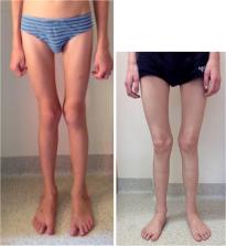
          Cystinosis is an autosomal recessive storage disease due to impaired transport of cystine out of lysosomes. Since the accumulation of intracellular cystine affects all organs and tissues, the management of cystinosis requires a specialized multidisciplinary team consisting of pediatricians, nephrologists, nutritionists, ophthalmologists, endocrinologists, neurologists' geneticists, and orthopedic surgeons. Treatment with cysteamine can delay or prevent most clinical manifestations of cystinosis, except the renal Fanconi syndrome. Virtually all individuals with classical, nephropathic cystinosis suffer from cystinosis metabolic bone disease (CMBD), related to the renal Fanconi syndrome in infancy and progressive chronic kidney disease (CKD) later in life. Manifestations of CMBD include hypophosphatemic rickets in infancy, and renal osteodystrophy associated with CKD resulting in bone deformities, osteomalacia, osteoporosis, fractures, and short stature. Assessment of CMBD involves monitoring growth, leg deformities, blood levels of phosphate, electrolytes, bicarbonate, calcium, and alkaline phosphatase, periodically obtaining bone radiographs, determining levels of critical hormones and vitamins, such as thyroid hormone, parathyroid hormone, 25(OH) vitamin D, and testosterone in males, and surveillance for nonrenal complications of cystinosis such as myopathy. Treatment includes replacement of urinary losses, cystine depletion with oral cysteamine, vitamin D, hormone replacement, physical therapy, and corrective orthopedic surgery. The recommendations in this article came from an expert meeting on CMBD that took place in Salzburg, Austria, in December 2016.

          Related collections

          Most cited references55

          • Record: found
          • Abstract: found
          • Article: not found

          A novel gene encoding an integral membrane protein is mutated in nephropathic cystinosis.

          Nephropathic cystinosis, an autosomal recessive disorder resulting from defective lysosomal transport of cystine, is the most common inherited cause of renal Fanconi syndrome. The cystinosis gene has been mapped to chromosome 17p13. We found that the locus D17S829 was homozygously deleted in 23 out of 70 patients, and identified a novel gene, CTNS, which mapped to the deletion interval. CTNS encodes an integral membrane protein, cystinosin, with features of a lysosomal membrane protein. Eleven different mutations, all predicted to cause loss of function of the protein, were found to segregate with the disorder.
            Bookmark
            • Record: found
            • Abstract: found
            • Article: not found

            Vitamin D status and mortality risk in CKD: a meta-analysis of prospective studies.

            Vitamin D deficiency, assessed as low 25-hydroxyvitamin D (25[OH]D) level, is highly prevalent in patients with chronic kidney disease (CKD) and is associated with various adverse health outcomes. Whether low 25(OH)D levels in patients with CKD are an independent risk factor for mortality remains to be studied in detail, and this was the objective of our work. A systematic review and meta-analysis of prospective observational studies. Patients with CKD. CKD was diagnosed mainly as decreased estimated glomerular filtration rate. We performed a systematic literature search in MEDLINE, ISI, and EMBASE to identify prospective studies reporting on 25(OH)D levels and mortality. 25(OH)D serum concentrations. All-cause mortality. 10 studies with an overall sample of 6,853 patients with CKD were included. Relative risk of mortality per 10-ng/mL (25-nmol/L) increase in 25(OH)D level was 0.86 (95% CI, 0.82-0.91), with no indication of publication bias or significant heterogeneity (I(2) =15%; P = 0.3). Summary estimates for CKD cohorts with and without dialysis treatment showed homogeneous results (P = 0.9). Results may be limited by heterogeneity, unconsidered confounders, and the observational design of the studies. Furthermore, publication bias by unpublished null findings on the association of 25(OH)D level and mortality cannot be ruled out and ascertainment of CKD was based largely on estimated glomerular filtration rate. Higher 25(OH)D levels are associated with significantly improved survival in patients with CKD. Whether treatment of low 25(OH)D level using natural vitamin D supplementation improves survival in patients with CKD remains to be elucidated in randomized controlled trials. Copyright © 2011 National Kidney Foundation, Inc. Published by Elsevier Inc. All rights reserved.
              Bookmark
              • Record: found
              • Abstract: found
              • Article: not found

              Normative data for iliac bone histomorphometry in growing children.

              Many insights into normal and pathologic bone development can only be gained by bone histomorphometry. However, the use of this technique in pediatrics has so far been hampered by the lack of reference data. Therefore, we obtained transfixing iliac bone samples from 58 individuals between 1.5 and 22.9 years of age (25 male; tetracycline labeling performed in 48 subjects), who underwent surgery for reasons independent of abnormalities in bone development and metabolism. The results of histomorphometric analyses of cancellous parameters and cortical width are presented as means and standard deviations, as well as medians and ranges in five age groups. In addition, the original data are available from the authors. There were significant age-dependent increases in both cortical width and cancellous bone volume, the latter being due to an increase in trabecular thickness. Osteoid thickness did not vary significantly with age. Bone surface-based indicators of bone formation showed an age-dependent decline, reflecting similar changes in activation frequency. Mineral apposition rate decreased continuously with age. Parameters of bone resorption did not vary significantly between age groups. Paired biopsies from adjacent sites, obtained in eight subjects, were used to examine the reproducibility of histomorphometric parameters in children. The lowest coefficients of variation (<10%) were found for structural measures, as well as mineral apposition rate and wall thickness. The highest variability was found for cellular parameters. The availability of reference material will greatly facilitate the use of histomorphometry in pediatrics.
                Bookmark

                Author and article information

                Contributors
                katharina.hohenfellner@ro-med.de
                Journal
                J Inherit Metab Dis
                J. Inherit. Metab. Dis
                10.1002/(ISSN)1573-2665
                JIMD
                Journal of Inherited Metabolic Disease
                John Wiley & Sons, Inc (Hoboken, USA )
                0141-8955
                1573-2665
                05 August 2019
                September 2019
                : 42
                : 5 ( doiID: 10.1002/jimd.v42.5 )
                : 1019-1029
                Affiliations
                [ 1 ] Ro Med Kliniken, Pediatric Nephrology Rosenheim Germany
                [ 2 ] Shriners Hospital for Children, McGill University Montreal Canada
                [ 3 ] Service of Pediatric Nephrology University Hospital Vall d’ Hebron Barcelona Spain
                [ 4 ] Department of Nephrology Children's University Hospital Dublin Ireland
                [ 5 ] Référence Center for Rare Renal Diseases Hôpital Femme‐Mère‐Enfant Bron France
                [ 6 ] Department of Medicine University Hospital Freiburg Freiburg Germany
                [ 7 ] Division of Pediatric Endocrinology Children's Hospital and Polyclinic iSPZ, Dr. v. Haunerschen Kinderspital, University Hospital Munich Munich Germany
                [ 8 ] Department of Orthopaedic Surgery Children's University Hospital Dublin Ireland
                [ 9 ] Department of Pediatric Nephrology Hôpital Robert‐Debré and University of Paris Diderot Paris France
                [ 10 ] Department of Pediatrics Baylor College of Medicine and Texas Children's Hospital Houston Texas
                [ 11 ] National Human Genome Research Institute National Institutes of Health Undiagnosed Diseases Program Bethesda Maryland
                [ 12 ] Department of Diagnostic and Interventional Radiology Klinikum Traunstein Traunstein Germany
                [ 13 ] Children‘s University Hospital Muenster Muenster Germany
                [ 14 ] Schoen Clinic Munich Harlaching Specialist Centre for Paediatric and Neuro‐Orthopaedics Munich Germany
                [ 15 ] Division of Pediatric Nephrology University Children's Hospital Bonn Germany
                [ 16 ] Kliniken Südostbayern AG, Sozialpädiatrisches Zentrum Traunstein Germany
                [ 17 ] Department of Pediatrics & Development and Regeneration University Hospitals Leuven & Katholieke Universiteit Leuven Leuven Belgium
                [ 18 ] National Institutes of Health, National Human Genome Research Institute (NHGRI) Bethesda Maryland
                [ 19 ] Hospital Universitario Central de Asturias Pediatría Oviedo Spain
                [ 20 ] Department of General Pediatrics University Children's Hospital Münster Münster Germany
                [ 21 ] Reference Center of Inherited Metabolic Diseases, Nephrology Unit, Hospital Necker Enfants Malades, APHP University Paris Descartes Paris France
                [ 22 ] Department of Pediatrics, Center of Pediatric Nephrology and Transplantation (CPNT), Kasr Al Ainy Faculty of Medicine Cairo University Cairo Egypt
                [ 23 ] Department of Pediatrics, Department of Pediatric Nephrology Ro‐Med Kliniken Rosenheim Germany
                [ 24 ] Department of Pediatric Nephrology Hacettepe University Faculty of Medicine Ankara Turkey
                [ 25 ] Department of Nephrology National Medical and Research Center for Children's Health Moscow Russia
                [ 26 ] Pediatric Clinic Wildermeth Children's Hospital Biel‐Bienne Switzerland
                [ 27 ] Institute of Osteology and Biomechanics University Medical Center Hamburg‐Eppendorf, University of Hamburg Hamburg Germany
                [ 28 ] Department of Pediatric Kidney, Liver and Metabolic Diseases Hannover Medical School Hannover Germany
                Author notes
                [*] [* ] Correspondence

                Katharina Hohenfellner, Abt. Kindernephrologie, Klinik für Kinder und Jugendheilkunde, Pettenkoferstrasse 10, 83022 Rosenheim, Germany.

                Email: katharina.hohenfellner@ 123456ro-med.de

                Author information
                https://orcid.org/0000-0003-0978-8087
                Article
                JIMD12134
                10.1002/jimd.12134
                7379238
                31177550
                87289727-f123-40e0-a03e-fe29f91bf0ae
                © 2019 The Authors. Journal of Inherited Metabolic Disease published by John Wiley & Sons Ltd on behalf of SSIEM

                This is an open access article under the terms of the http://creativecommons.org/licenses/by-nc-nd/4.0/ License, which permits use and distribution in any medium, provided the original work is properly cited, the use is non‐commercial and no modifications or adaptations are made.

                History
                : 25 April 2019
                : 02 June 2019
                : 03 June 2019
                Page count
                Figures: 4, Tables: 3, Pages: 11, Words: 6953
                Funding
                Funded by: Cystinosis Foundation Germany
                Funded by: Cystinosis Foundation , open-funder-registry 10.13039/100003719;
                Categories
                Original Article
                Original Articles
                Custom metadata
                2.0
                September 2019
                Converter:WILEY_ML3GV2_TO_JATSPMC version:5.8.5 mode:remove_FC converted:24.07.2020

                Internal medicine
                chronic kidney disease,ckd‐mbd,cystinosis,cystinosis metabolic bone disease,fanconi syndrome,hypophosphatemic rickets,transplantation

                Comments

                Comment on this article