14
views
0
recommends
+1 Recommend
0 collections
    0
    shares
      • Record: found
      • Abstract: found
      • Article: found
      Is Open Access

      Increasing fetal ovine number per gestation alters fetal plasma clinical chemistry values

      research-article

      Read this article at

      Bookmark
          There is no author summary for this article yet. Authors can add summaries to their articles on ScienceOpen to make them more accessible to a non-specialist audience.

          Abstract

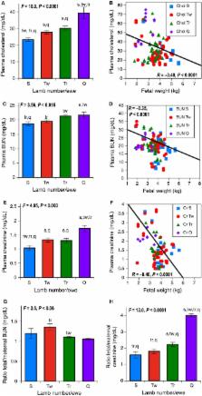
          Intrauterine growth restriction ( IUGR) is interconnected with developmental programming of lifelong pathophysiology. IUGR is seen in human multifetal pregnancies, with stepwise rises in fetal numbers interfering with placental nutrient delivery. It remains unknown whether fetal blood analyses would reflect fetal nutrition, liver, and excretory function in the last trimester of human or ovine IUGR. In an ovine model, we hypothesized that fetal plasma biochemical values would reflect progressive placental, fetal liver, and fetal kidney dysfunction as the number of fetuses per gestation rose. To determine fetal plasma biochemical values in singleton, twin, triplet, and quadruplet/quintuplet ovine gestation, we investigated morphometric measures and comprehensive metabolic panels with nutritional measures, liver enzymes, and placental and fetal kidney excretory measures at gestational day ( GD) 130 (90% gestation). As anticipated, placental dysfunction was supported by a stepwise fall in fetal weight, fetal plasma glucose, and triglyceride levels as fetal number per ewe rose. Fetal glucose and triglycerides were directly related to fetal weight. Plasma creatinine, reflecting fetal renal excretory function, and plasma cholesterol, reflecting placental excretory function, were inversely correlated with fetal weight. Progressive biochemical disturbances and growth restriction accompanied the rise in fetal number. Understanding the compensatory and adaptive responses of growth‐restricted fetuses at the biochemical level may help explain how metabolic pathways in growth restriction can be predetermined at birth. This physiological understanding is important for clinical care and generating interventional strategies to prevent altered developmental programming in multifetal gestation.

          Related collections

          Most cited references28

          • Record: found
          • Abstract: found
          • Article: not found

          The developmental origins of adult disease (Barker) hypothesis.

          Many studies have provided evidence for the hypothesis that size at birth is related to the risk of developing disease in later life. In particular, links are well established between reduced birthweight and increased risk of coronary heart disease, diabetes, hypertension and stroke in adulthood. These relationships are modified by patterns of postnatal growth. The most widely accepted mechanisms thought to underlie these relationships are those of fetal programming by nutritional stimuli or excess fetal glucocorticoid exposure. It is suggested that the fetus makes physiological adaptations in response to changes in its environment to prepare itself for postnatal life. These changes may include epigenetic modification of gene expression. Less clear at this time are the relevance of fetal programming phenomena to twins and preterm babies, and whether any of these effects can be reversed after birth. Much current active research in this field will be of direct relevance to future obstetric practice.
            Bookmark
            • Record: found
            • Abstract: found
            • Article: not found

            Renal physiology of pregnancy.

            Pregnancy involves remarkable orchestration of physiologic changes. The kidneys are central players in the evolving hormonal milieu of pregnancy, responding and contributing to the changes in the environment for the pregnant woman and fetus. The functional impact of pregnancy on kidney physiology is widespread, involving practically all aspects of kidney function. The glomerular filtration rate increases 50% with subsequent decrease in serum creatinine, urea, and uric acid values. The threshold for thirst and antidiuretic hormone secretion are depressed, resulting in lower osmolality and serum sodium levels. Blood pressure drops approximately 10 mmHg by the second trimester despite a gain in intravascular volume of 30% to 50%. The drop in systemic vascular resistance is multifactorial, attributed in part to insensitivity to vasoactive hormones, and leads to activation of the renin-aldosterone-angiostensin system. A rise in serum aldosterone results in a net gain of approximately 1000 mg of sodium. A parallel rise in progesterone protects the pregnant woman from hypokalemia. The kidneys increase in length and volume, and physiologic hydronephrosis occurs in up to 80% of women. This review will provide an understanding of these important changes in kidney physiology during pregnancy, which is fundamental in caring for the pregnant patient.
              Bookmark
              • Record: found
              • Abstract: found
              • Article: not found

              The effect of intrauterine growth retardation on the development of renal nephrons.

              To investigate the effect of Type II (asymmetrical) intrauterine growth retardation (IUGR) on renal development. A prospective descriptive study. Department of Fetal and Infant Pathology, Liverpool Children's Hospital. Six (severely) affected IUGR stillbirths of known gestational age with a control group of stillbirths with birthweight greater than 10th centile, and eight liveborn IUGR infants who died within a year of birth with a control group of appropriately grown infants who died within a year of birth (postnatal groups). The kidneys from all the groups studied were analysed using unbiased, reproducible and objective design-based stereological techniques. Total renal nephron (glomerular) numbers and average volumes of total nephron and cortical and medullary nephron segments. Nephron number estimates lay below the control group's 5% prediction limit in five out of the six growth-retarded stillbirths, and were significantly (P less than 0.005, IUGR at 65% of the control mean) reduced in the postnatal group. Estimates of nephron (segment) volume did not differ between control and IUGR groups. Type II intrauterine growth retardation may exert a profound effect on renal development. The reduced nephron number at birth, together with the lack of any early postnatal compensation in either nephron number or nephron size, emphasizes the need for vigorous antenatal surveillance for IUGR and consideration of elective preterm delivery of affected fetuses. A systematic review of other organs, which develop in a similarly rapid fashion during the late intrauterine period, is indicated by this work. With one exception, all birthweights in the growth-retarded groups were below the third centile, thus the precise quantitative relation between progressive IUGR and renal function requires further assessment.
                Bookmark

                Author and article information

                Journal
                Physiol Rep
                Physiol Rep
                10.1002/(ISSN)2051-817X
                PHY2
                physreports
                Physiological Reports
                John Wiley and Sons Inc. (Hoboken )
                2051-817X
                26 August 2016
                August 2016
                : 4
                : 16 ( doiID: 10.1111/phy2.2016.4.issue-16 )
                : e12905
                Affiliations
                [ 1 ] Departments of PediatricsUniversity of Wisconsin‐Madison School of Medicine and Public Health Madison WIUSA
                [ 2 ] Obstetrics and Gynecology Perinatal Research LaboratoriesUniversity of Wisconsin‐Madison School of Medicine and Public Health Madison WIUSA
                [ 3 ] Department of PediatricsUniversity of Iowa Children's Hospital Iowa IAUSA
                Author notes
                [*] [* ] Correspondence

                Pamela J. Kling, Department of Pediatrics, Meriter Hospital, University of Wisconsin, 202 S. Park St., Madison, WI 53715, USA.

                Tel: +608 417‐6236

                Fax: +608 417‐6377

                E‐mail: pkling@ 123456pediatrics.wisc.edu

                Article
                PHY212905
                10.14814/phy2.12905
                5002913
                27565903
                87ae9885-79b0-4f32-bf12-e410a6dd20da
                © 2016 The Authors. Physiological Reports published by Wiley Periodicals, Inc. on behalf of the American Physiological Society and The Physiological Society.

                This is an open access article under the terms of the Creative Commons Attribution License, which permits use, distribution and reproduction in any medium, provided the original work is properly cited.

                History
                : 08 June 2016
                : 22 July 2016
                : 27 July 2016
                Page count
                Pages: 12
                Funding
                Funded by: Hilldale Undergraduate/Faculty Research Fellowship
                Funded by: UW Department of Pediatrics
                Funded by: NIH
                Award ID: HL087144
                Award ID: HL079020
                Award ID: P01 HD38843
                Funded by: Meriter Foundation
                Categories
                Nutrition
                Reproductive Conditions, Disorders and Treatments
                Maternal, Fetal and Neonatal Physiology
                Metabolic Pathways
                Original Research
                Original Research
                Custom metadata
                2.0
                phy212905
                August 2016
                Converter:WILEY_ML3GV2_TO_NLMPMC version:4.9.4 mode:remove_FC converted:29.08.2016

                fetal,nutrition,ovine,placenta
                fetal, nutrition, ovine, placenta

                Comments

                Comment on this article