- Record: found
- Abstract: found
- Article: found
Immobile Tricuspid Valve: Incidental Finding in a Case of Terminal Cardiomyopathy Due to Thalassemia Major
case-report
There is no author summary for this article yet. Authors can add summaries to their articles on ScienceOpen to make them more accessible to a non-specialist audience.
Abstract
Introduction
Thalassemia Major is an inherited disorder caused by impaired synthesis of the B
globin chain and characterized by ineffective erythropoiesis that requires regular,
lifelong transfusion therapy, which creates a state of iron overload.
1
Once reticuloendothelial stores
saturate, iron deposition increases in myocardium such as other parenchymal
tissues.
2
Cardiac
complications due to this deposition are the leading cause of death. After a silent
first decade, iron deposits in the cardiac tissue lead to arrhythmias, systolic and
diastolic dysfunction, and congestive heart failure in the second or third
decade.
3
In this case
report, we present an adolescent girl who did not receive regular iron chelation
therapy and had cardiomyopathy, arrhythmia and immobile tricuspid valve secondary
to
thalassemia major.
Case presentation
A 14-year-old Syrian girl with Thalassemia Major presented to the emergency room
with a three-month history of increasing fatigue, dyspnea, and abdominal
distension. Her medical history revealed that she had been diagnosed with
Thalassemia Major at the age of one year old, and she received irregular
erythrocyte transfusion and iron chelation therapy in her country. It was
learned that the compliance for previous blood transfusion and chelation therapy
was very poor. On general examination, she was undernourished with short stature
(body weight < 25 p, height < 3p) and the physical examination revealed
dyspnea with a typical facial thalassemic feature without cyanosis.
Chest x-ray showed areas of consolidation on both sides of the lungs and
increased cardiothoracic ratio (Figure 1).
The electrocardiogram showed sinus rhythm with 70/min heart rate and
prolongation of QTc value with 0.46 seconds (Figure 2-A). Transthoracic echocardiography
revealed both ventricle
systolic and diastolic ventricular dysfunction, left ventricle ejection fraction
was 48% and fractional shortening was 24% were calculated with a mild left
ventricle dilatation (Table 1).
Mild-moderate mitral regurgitation and trivial pericardial effusion were also
observed. Right ventricular inflow view in systole showing thickened, immobile
leaflets of tricuspid valve in a fixed open position, causing mal-coaptation and
severe regurgitation without stenosis (see Figure
3 and Video 1). Apical
four-chamber view in diastole showed immobile leaflets of tricuspid valve in a
fixed open position, as showed by the color Doppler (Video 2) (See additional files
Video 3, 4 and 5). Right atrial, right ventricle dilatation
and minimal pulmonary regurgitation with mild pulmonary hypertension were also
observed.
Figure 1
Chest X-Ray of the patient.
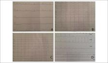
Figure 2
Electrocardiography of the patient. (A). Sinus rhythm with QTc
prolongation (B). Atrial flutter (C). Atrioventricular dissociation
and ventricular extra-systole (D). Holter monitorization revealed
non-sustained ventricular tachycardia.
Table 1
Echocardiographic measurements of the patient
Data
Values
M-Mode Measurements
LVID, cm
4.9
Ejection Fraction
48
Fractional shortening, %
24
RVID, cm
4.8
Doppler Measurements
Tricuspid E, cm/s
81
Tricuspid A, cm/s
25
Tricuspid E/A
3.2
Tissue Doppler Measurements (RV)
E' cm/s
12.1
A' cm/s
7.8
E'/A'
1.55
E/E'
6.7
S'
11.4
IVCT, ms
65
IVRT, ms
78
RV MPI
62
A: peak late diastolic velocity; A': late diastolic velocity; E: peak
early diastolic velocity; E': early diastolic velocity; ET: ejection
time; IVCT: isovolumic contraction time; IVRT: isovolumic relation
time; LVIDd: Left ventricular internal diastolic diameter; MPI:
myocardial performance index; RV: right ventricle; RVID: right
ventricular internal diameter; S': systolic velocity; Tissue Doppler
imaging of the tricuspid valve.
Figure 3
Transthoracic echocardiography from the parasternal view by tilting
transducer inferomedially exploring the right atrium (RA) and right
ventricle (RV) inflow tract; immobile leaflets of the tricuspid
valve (TV) leading to severe insufficiency.
Video 1
Transthoracic echocardiography from the right ventricular inflow view
in systole showing immobile leaflets of tricuspid valve in a fixed
open position, causing mal-coaptation and severe regurgitation. To
view the video click on the link: https://bit.ly/2lTc6iX
Video 2
Transthoracic echocardiography from the apical four-chamber by color
Doppler view showed immobile leaflets of tricuspid valve in a fixed
open position. To view the video click on the link: https://bit.ly/2lTc6iX
Video 3
Transthoracic echocardiography from the parasternal long axis view
with color Doppler showing mild mitral regurgitation. To view the
video click on the link: https://bit.ly/2lTc6iX
Video 4
Transthoracic echocardiography from the parasternal long axis view
showing normal systolic function of the left ventricle and minimal
pericardial effusion. To view the video click on the link: https://bit.ly/2lTc6iX
Video 5
Transthoracic echocardiography from the parastrenal short axis view
showed enlargement of the right ventricle and pericardial effusion.
To view the video click on the link: https://bit.ly/2lTc6iX
After hospitalization in the intensive care unit, inotropes, diuretics and iron
chelation treatment (Dopamine, Dobutamine, Furosemide infusion, Propranolol,
Enalapril, Aldactone and Deferoxamine, Deferiprone therapy) started as soon as
possible. Cardiac enzymes were sent to screen possible myocarditis, and D-dimer
was sent to detect pulmonary thromboembolism. Results were found to be negative.
On the seventh day of hospitalization, the electrocardiogram showed atrial
flutter (Figure 2-B). Therefore, digoxin
and low molecular weight heparin treatment were also started. In the second
week, the patient developed acute renal insufficiency and the electrocardiogram
showed atrioventricular dissociation and ventricular extra-systole (Figure 2-C). Immediately
after the digoxin
treatment had been stopped, amiodarone infusion has started. Holter
monitorization revealed atrioventricular dissociation and non-sustained
ventricular tachycardia (Figure 2-D). Blood
level of Digoxin was within normal reference values. Serial echocardiography was
performed and no difference has been observed in the cardiac parameters during
the hospitalization. Despite atrioventricular dissociation, we decided to
follow-up her without pacemaker implantation due to hemodynamic stability.
Although secondary prevention of implantable cardioverter defibrillator was
decided after the patient was taken to control ventricular arrhythmias with
amiodarone, she died due to ventricular tachycardia on the 22nd day of
hospitalization.
Discussion
In thalassemia, cardiovascular system involvement is pivotal in the prognosis and
quality of life. Iron overload cardiomyopathy is the leading cause of mortality
accounts up to 67% and 71% in thalassemia.
4
As iron overload, multiple factors such as chronic anemia,
hypersplenism, non-progressive restrictive lung disease also lead to cardiac
complications in Thalassemia Major.
5
Iron is mainly stored in myocytes and other cells in the form of
free iron, also ferritin and hemosiderin. Free iron, which is referred to as labile
cellular iron, is the most toxic form of iron and also the most accessible form for
chelation. The goal of iron chelation therapy is to reduce the iron deposition
especially in plasma and other tissues. In some cases, these heart complications
were reported as reversible with early detection of iron overload and response to
regular iron chelation therapy.
6
Cardiac magnetic resonance imaging (MRI) is the gold standard for detecting
myocardial iron deposition. In our case, cardiac MRI was not performed due to lack
of experienced staff in our hospital. Progressive increase of brain natriuretic
peptide assay is highly sensitive and specific in the diagnosis of heart failure.
In
our patient, brain natriuretic peptide levels were markedly elevated.
Conventional standard echocardiography exhibits pathologic findings at advanced
stages of cardiac involvement. The assessment of the ventricular function involves
two different phenotypes. The first one is ‘dilated cardiomyopathy’ phenotype
revealed by with left-right ventricular dilatation and reduced contractility, which
cause congestive heart failure. The second one is ‘restrictive cardiomyopathy’
phenotype revealed by restrictive left-right ventricular filling resulting in
pulmonary hypertension, right ventricular dilatation, and heart failure.
7
In this report, our patient had impaired cardiac functions similar to both dilated
and restrictive patterns of cardiomyopathy. Right, and left ventricle contractility
was reduced which led to congestive heart failure. Both ventricle diastolic
dimensions were increased. Assessment with pulsed and pulsed tissue Doppler
demonstrated left and right ventricle diastolic dysfunction. Factors that may cause
pulmonary hypertension in patients with thalassemia include elevated pulmonary
resistance due to high volume of blood flow, elevated shear forces, hypercoagulable
state secondary to splenectomy and nitric oxide formation after chronic hemolysis.
Although right heart failure may develop secondary to pulmonary hypertension, in
thalassemic patients, it may also develop in the absence of elevated pulmonary
hypertension.
8
In our case, typical stenotic changes and doming that seen in rheumatic diseases were
not present in the tricuspid leaflet. Uniformly, mildly thickened tricuspid leaflets
were present with a relatively fixed valve orifice without stenosis. Cardiac
carcinoid usually affects the right cardiac chamber of the heart and results in a
similar presentation. However, it is not reported in the pediatric age group in the
literature. Nevertheless, carcinoid tumor should also be considered as a
differential diagnosis in isolated advanced tricuspid valve involvement.
9
In our case in contrast to the
carcinoid tumor, the tricuspid valve did not exhibit very bright echoes secondary
to
fibrous plaques that are deposited on the endocardium of the leaflets.
10
Biogenic amine levels in plasma
and urine samples were found to be in the normal range that excluded diagnosis of
carcinoid tumor. The related literature indicates similar findings in patients with
thalassemia; however, illustration of echocardiograms in children is not
satisfactory. Aessopos et al.
6
reported valvular involvement including leaflet thickening (48%), endocardial
calcification (20%), and left-sided valve regurgitation in adult patients with
thalassemia intermedia.
6
In our
case, the patient had serious dysrhythmias due to endocardial involvement,
contraction and relaxation dysfunction due to myocardial involvement, and severe
leaflet disorder due to valvular involvement. We herein report an extraordinary
thalassemia major patient with immobile and non-stenotic tricuspid valve that
emerges as a part of the terminal phase of the cardiomyopathy.
Conclusion
Thalassemia major patients, especially those who do not receive regular chelation
therapy, are under great risk of cardiac involvement. Early detection and regular
treatment regimen enhance their survival and quality of life. We firstly present an
immobile tricuspid valve in an adolescent girl. This very rare case of severe
cardiac findings due to iron deposition is associated with endocardial, myocardial
and valvular involvement. In patients with thalassemia, these end-stage
complications of the cardiovascular system are irreversible despite treatment.
Related collections
Most cited references6
- Record: found
- Abstract: found
- Article: not found
Beta-thalassemia cardiomyopathy: history, present considerations, and future perspectives.
- Record: found
- Abstract: found
- Article: not found
Cardiac involvement in thalassemia intermedia: a multicenter study.
D Loukopoulos, J Joussef, M Karagiorga … (2001)
- Record: found
- Abstract: found
- Article: not found
Cardiac complications in thalassemia major.
John Wood (2009)