4
views
0
recommends
+1 Recommend
0 collections
    0
    shares
      • Record: found
      • Abstract: found
      • Article: found
      Is Open Access

      microRNA‐148a‐3p in extracellular vesicles derived from bone marrow mesenchymal stem cells suppresses SMURF1 to prevent osteonecrosis of femoral head

      research-article

      Read this article at

      Bookmark
          There is no author summary for this article yet. Authors can add summaries to their articles on ScienceOpen to make them more accessible to a non-specialist audience.

          Abstract

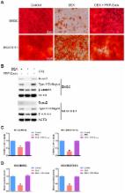
          Extracellular vesicle (EV)‐associated microRNAs (miRNAs) have been found as the important biomarkers participating in the development of osteonecrosis of the femoral head (ONFH). Consequently, this study sought to examine the underlying mechanism of bone marrow mesenchymal stem cell (BMSC)‐derived EVs containing miR‐148a‐3p in ONFH. The ONFH rat models were established. Reverse transcription quantitative polymerase chain reaction (RT‐qPCR) and Western blot analysis were applied to detect miR‐148a‐3p, Smad ubiquitination regulatory factor 1 (SMURF1), SMAD7 and B‐cell CLL/lymphoma 2 (BCL2) expression, followed by determination of relationship between miR‐148a‐3p and SMURF1. BMSCs were isolated from normal rats and ONFH rats, and EVs were extracted from BMSCs of normal rats. BMSCs from ONFH rats were treated with mimic, inhibitor, small interfering RNA or EVs from miR‐148a‐3p mimic‐treated BMSCs from normal rats (BMSC‐EV‐miR‐148a‐3p mimic). Cell Counting Kit‐8 and alizarin red staining were utilized to detect cell viability and osteogenic differentiation of BMSCs. ONFH rats were injected with BMSC‐EV‐miR‐148a‐3p mimic to explore the function of BMSC‐EV‐delivered miR‐148a‐3p in vivo. miR‐148a‐3p was down‐regulated in BMSCs and EVs from ONFH rats following decreased BMSCs viability and osteogenic differentiation. SMURF1 was a target gene of miR‐148a‐3p, and resulted in ubiquitination and degradation of SMAD7 to decreased BCL2 expression. The proliferation and differentiation of BMSCs were promoted by BMSC‐EV‐miR‐148a‐3p mimic or SMURF1 silencing. Additionally, BMSC‐EV‐miR‐148a‐3p mimic increased cell proliferation and osteogenic response, diminished SMURF1 expression, and elevated SMAD7 and BCL2 expression in ONFH rats. Collectively, miR‐148a‐3p overexpressed in BMSC‐EVs promoted SMAD7 and BCL2 expression by inhibiting SMURF1, thus alleviating ONFH.

          Related collections

          Most cited references43

          • Record: found
          • Abstract: found
          • Article: found
          Is Open Access

          Mesenchymal Stem Cell‐Derived Extracellular Vesicles as Mediators of Anti‐Inflammatory Effects: Endorsement of Macrophage Polarization

          Abstract Mesenchymal Stem Cells (MSCs) are effective therapeutic agents enhancing the repair of injured tissues mostly through their paracrine activity. Increasing evidences show that besides the secretion of soluble molecules, the release of extracellular vesicles (EVs) represents an alternative mechanism adopted by MSCs. Since macrophages are essential contributors toward the resolution of inflammation, which has emerged as a finely orchestrated process, the aim of the present study was to carry out a detailed characterization of EVs released by human adipose derived‐MSCs to investigate their involvement as modulators of MSC anti‐inflammatory effects inducing macrophage polarization. The EV‐isolation method was based on repeated ultracentrifugations of the medium conditioned by MSC exposed to normoxic or hypoxic conditions (EVNormo and EVHypo). Both types of EVs were efficiently internalized by responding bone marrow‐derived macrophages, eliciting their switch from a M1 to a M2 phenotype. In vivo, following cardiotoxin‐induced skeletal muscle damage, EVNormo and EVHypo interacted with macrophages recruited during the initial inflammatory response. In injured and EV‐treated muscles, a downregulation of IL6 and the early marker of innate and classical activation Nos2 were concurrent to a significant upregulation of Arg1 and Ym1, late markers of alternative activation, as well as an increased percentage of infiltrating CD206pos cells. These effects, accompanied by an accelerated expression of the myogenic markers Pax7, MyoD, and eMyhc, were even greater following EVHypo administration. Collectively, these data indicate that MSC‐EVs possess effective anti‐inflammatory properties, making them potential therapeutic agents more handy and safe than MSCs. stem cells translational medicine 2017 Stem Cells Translational Medicine 2017;6:1018–1028
            Bookmark
            • Record: found
            • Abstract: found
            • Article: found
            Is Open Access

            Extracellular Vesicles Derived from Bone Marrow Mesenchymal Stem Cells Protect against Experimental Colitis via Attenuating Colon Inflammation, Oxidative Stress and Apoptosis

            The administration of bone mesenchymal stem cells (BMSCs) could reverse experimental colitis, and the predominant mechanism in tissue repair seems to be related to their paracrine activity. BMSCs derived extracellular vesicles (BMSC-EVs), including mcirovesicles and exosomes, containing diverse proteins, mRNAs and micro-RNAs, mediating various biological functions, might be a main paracrine mechanism for stem cell to injured cell communication. We aimed to investigate the potential alleviating effects of BMSC-EVs in 2,4,6-trinitrobenzene sulfonic acid (TNBS)-induced colitis model. Intravenous injection of BMSC-EVs attenuated the severity of colitis as evidenced by decrease of disease activity index (DAI) and histological colonic damage. In inflammation response, the BMSC-EVs treatment significantly reduced both the mRNA and protein levels of nuclear factor kappaBp65 (NF-κBp65), tumor necrosis factor-alpha (TNF-α), induciblenitric oxidesynthase (iNOS) and cyclooxygenase-2 (COX-2) in injured colon. Additionally, the BMSC-EVs injection resulted in a markedly decrease in interleukin-1β (IL-1β) and an increase in interleukin-10 (IL-10) expression. Therapeutic effect of BMSC-EVs associated with suppression of oxidative perturbations was manifested by a decrease in the activity of myeloperoxidase (MPO) and Malondialdehyde (MDA), as well as an increase in superoxide dismutase (SOD) and glutathione (GSH). BMSC-EVs also suppressed the apoptosis via reducing the cleavage of caspase-3, caspase-8 and caspase-9 in colitis rats. Data obtained indicated that the beneficial effects of BMSC-EVs were due to the down regulation of pro-inflammatory cytokines levels, inhibition of NF-κBp65 signal transduction pathways, modulation of anti-oxidant/ oxidant balance, and moderation of the occurrence of apoptosis.
              Bookmark
              • Record: found
              • Abstract: found
              • Article: found
              Is Open Access

              Exosomes derived from human platelet-rich plasma prevent apoptosis induced by glucocorticoid-associated endoplasmic reticulum stress in rat osteonecrosis of the femoral head via the Akt/Bad/Bcl-2 signal pathway

              An excess of glucocorticoids (GCs) is reported to be one of the most common causes of osteonecrosis of the femoral head (ONFH). In addition, GCs can induce bone cell apoptosis through modulating endoplasmic reticulum (ER) stress. Among the three main signal pathways in ER stress, the PERK (protein kinase RNA-like ER kinase)/CHOP (CCAAT-enhancer-binding protein homologous protein) pathway has been considered to be closely associated with apoptosis. Platelet-rich plasma (PRP) has been referred to as a concentration of growth factors and the exosomes derived from PRP (PRP-Exos) have a similar effect to their parent material. The enriched growth factors can be encapsulated into PRP-Exos and activate Akt and Erk pathways to promote angiogenesis. Activation of the Akt pathway may promote the expression of anti-apoptotic proteins like Bcl-2, while CHOP can inhibit B-cell lymphoma 2 (Bcl-2) expression to increase the level of cleaved caspase-3 and lead to cell death. Consequently, we hypothesized that PRP-Exos prevent apoptosis induced by glucocorticoid-associated ER stress in rat ONFH via the Akt/Bad/Bcl-2 signal pathway. To verify this hypothesis, a dexamethasone (DEX)-treated in vitro cell model and methylprednisolone (MPS)-treated in vivo rat model were adopted. Characterization of PRP-Exos, and effects of PRP-Exos on proliferation, apoptosis, angiogenesis, and osteogenesis of cells treated with GCs in vitro and in vivo were examined. Furthermore, the mechanism by which PRP-Exos rescue the GC-induced apoptosis through the Akt/Bad/Bcl-2 pathway was also investigated. The results indicate that PRP-Exos have the capability to prevent GC-induced apoptosis in a rat model of ONFH by promoting Bcl-2 expression via the Akt/Bad/Bcl-2 signal pathway under ER stress.
                Bookmark

                Author and article information

                Contributors
                ningbo_duan@163.com
                Journal
                J Cell Mol Med
                J Cell Mol Med
                10.1111/(ISSN)1582-4934
                JCMM
                Journal of Cellular and Molecular Medicine
                John Wiley and Sons Inc. (Hoboken )
                1582-1838
                1582-4934
                01 September 2020
                October 2020
                : 24
                : 19 ( doiID: 10.1111/jcmm.v24.19 )
                : 11512-11523
                Affiliations
                [ 1 ] Department of Orthopedics Hunan Children's Hospital Changsha China
                [ 2 ] Department of Rehabilitation Xiangya Hospital of Central South University Changsha China
                [ 3 ] Department of Orthopedics Xiangya Hospital of Central South University Changsha China
                Author notes
                [*] [* ] Correspondence

                Ningbo Duan, Department of Rehabilitation, Xiangya Hospital of Central South University, No. 87, Xiangya Road, Kaifu District, Changsha 410008, Hunan Province, China.

                Email: ningbo_duan@ 123456163.com

                Author information
                https://orcid.org/0000-0002-8327-1660
                https://orcid.org/0000-0002-8723-0938
                https://orcid.org/0000-0002-9280-887X
                https://orcid.org/0000-0002-4666-6911
                https://orcid.org/0000-0001-5110-6313
                https://orcid.org/0000-0002-3986-5189
                https://orcid.org/0000-0002-8425-4088
                Article
                JCMM15766
                10.1111/jcmm.15766
                7576243
                32871042
                886f720c-d178-45fd-8fed-499daa74d7ab
                © 2020 The Authors. Journal of Cellular and Molecular Medicine published by Foundation for Cellular and Molecular Medicine and John Wiley & Sons Ltd.

                This is an open access article under the terms of the http://creativecommons.org/licenses/by/4.0/ License, which permits use, distribution and reproduction in any medium, provided the original work is properly cited.

                History
                : 29 April 2020
                : 22 July 2020
                : 03 August 2020
                Page count
                Figures: 4, Tables: 1, Pages: 12, Words: 7026
                Categories
                Original Article
                Original Articles
                Custom metadata
                2.0
                October 2020
                Converter:WILEY_ML3GV2_TO_JATSPMC version:5.9.3 mode:remove_FC converted:21.10.2020

                Molecular medicine
                bcl2,bone marrow mesenchymal stem cells,extracellular vesicles,microrna‐148a‐3p,osteonecrosis of the femeral head,smad7,smurf1

                Comments

                Comment on this article