11
views
0
recommends
+1 Recommend
0 collections
    0
    shares
      • Record: found
      • Abstract: found
      • Article: found
      Is Open Access

      Changing muscle function with sustained glial derived neurotrophic factor treatment of rabbit extraocular muscle

      research-article
      1 , 1 , 1 , 2 , *
      PLoS ONE
      Public Library of Science

      Read this article at

      Bookmark
          There is no author summary for this article yet. Authors can add summaries to their articles on ScienceOpen to make them more accessible to a non-specialist audience.

          Abstract

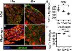
          Recent microarray and RNAseq experiments provided evidence that glial derived neurotrophic factor (GDNF) levels were decreased in extraocular muscles from human strabismic subjects compared to age-matched controls. We assessed the effect of sustained GDNF treatment of the superior rectus muscles of rabbits on their physiological and morphological characteristics, and these were compared to naïve control muscles. Superior rectus muscles of rabbits were implanted with a sustained release pellet of GDNF to deliver 2μg/day, with the contralateral side receiving a placebo pellet. After one month, the muscles were assessed using in vitro physiological methods. The muscles were examined histologically for alteration in fiber size, myosin expression patterns, neuromuscular junction size, and stem cell numbers and compared to age-matched naïve control muscles. GDNF resulted in decreased force generation, which was also seen on the untreated contralateral superior rectus muscles. Muscle relaxation times were increased in the GDNF treated muscles. Myofiber mean cross-sectional areas were increased after the GDNF treatment, but there was a compensatory increase in expression of developmental, neonatal, and slow tonic myosin heavy chain isoforms. In addition, in the GDNF treated muscles there was a large increase in Pitx2-positive myogenic precursor cells. One month of GDNF resulted in significant extraocular muscle adaptation. These changes are interesting relative to the decreased levels of GDNF in the muscles from subjects with strabismus and preliminary data in infant non-human primates where sustained GDNF treatment produced a strabismus. These data support the view that GDNF has the potential for improving eye alignment in subjects with strabismus.

          Related collections

          Most cited references47

          • Record: found
          • Abstract: found
          • Article: found
          Is Open Access

          Pervasive satellite cell contribution to uninjured adult muscle fibers

          Background Adult skeletal muscle adapts to functional needs, maintaining consistent numbers of myonuclei and stem cells. Although resident muscle stem cells or satellite cells are required for muscle growth and repair, in uninjured muscle, these cells appear quiescent and metabolically inactive. To investigate the satellite cell contribution to myofibers in adult uninjured skeletal muscle, we labeled satellite cells by inducing a recombination of LSL-tdTomato in Pax7CreER mice and scoring tdTomato+ myofibers as an indicator of satellite cell fusion. Results Satellite cell fusion into myofibers plateaus postnatally between 8 and 12 weeks of age, reaching a steady state in hindlimb muscles, but in extra ocular or diaphragm muscles, satellite cell fusion is maintained at postnatal levels irrespective of the age assayed. Upon recombination and following a 2-week chase in 6-month-old mice, tdTomato-labeled satellite cells fused into myofibers as 20, 50, and 80 % of hindlimb, extra ocular, and diaphragm myofibers, respectively, were tdTomato+. Satellite cells contribute to uninjured myofibers either following a cell division or directly without an intervening cell division. Conclusions The frequency of satellite cell fusion into the skeletal muscle fibers is greater than previously estimated, suggesting an important functional role for satellite cell fusion into adult myofibers and a requirement for active maintenance of satellite cell numbers in uninjured skeletal muscle.
            Bookmark
            • Record: found
            • Abstract: found
            • Article: found
            Is Open Access

            Treadmill Exercise Induced Functional Recovery after Peripheral Nerve Repair Is Associated with Increased Levels of Neurotrophic Factors

            Benefits of exercise on nerve regeneration and functional recovery have been reported in both central and peripheral nervous system disease models. However, underlying molecular mechanisms of enhanced regeneration and improved functional outcomes are less understood. We used a peripheral nerve regeneration model that has a good correlation between functional outcomes and number of motor axons that regenerate to evaluate the impact of treadmill exercise. In this model, the median nerve was transected and repaired while the ulnar nerve was transected and prevented from regeneration. Daily treadmill exercise resulted in faster recovery of the forelimb grip function as evaluated by grip power and inverted holding test. Daily exercise also resulted in better regeneration as evaluated by recovery of compound motor action potentials, higher number of axons in the median nerve and larger myofiber size in target muscles. Furthermore, these observations correlated with higher levels of neurotrophic factors, glial derived neurotrophic factor (GDNF), brain derived neurotrophic factor (BDNF) and insulin-like growth factor-1 (IGF-1), in serum, nerve and muscle suggesting that increase in muscle derived neurotrophic factors may be responsible for improved regeneration.
              Bookmark
              • Record: found
              • Abstract: found
              • Article: not found

              Cell adhesion and homeostatic synaptic plasticity.

              At synapses, pre- and post-synaptic cells get in direct contact with each other via cell adhesion molecules (CAMs). Several CAMs have been identified at the neuromuscular junction and at central synapses, where they regulate synaptic strength, by recruiting scaffolding proteins, neurotransmitter receptors and synaptic vesicles in response to the binding of counter-receptors across the synaptic cleft. Many synapses are also surrounded by astrocytic processes and embedded in conspicuous extracellular matrix (ECM). It is now widely recognized that astrocytes play a central role in regulating the synaptic machinery by exchanging information with the neuronal elements via diffusible molecules and direct physical interactions; this has lead to the concept of the 'tri-partite synapse'. More recently, the term 'tetra-partite synapse' has been introduced to underlie the importance of ECM in shaping synaptic function by mediating interaction and signaling between neurons and astrocytes. Here, we will review how this integrated view of the synapse can help us understand homeostatic synaptic plasticity at the neuromuscular junction and in the central nervous system. We will explore how synaptic CAMs regulate two forms of homeostatic plasticity: (i) postsynaptic scaling of synaptic currents to counteract changes in neuronal network activity and (ii) the compensatory modulation of presynaptic neurotransmitter release in response to changes in postsynaptic efficacy. We will discuss recent findings on activity-dependent trans-synaptic signaling events and the role of cell adhesion in the feedback control of network activity. This article is part of the Special Issue entitled 'Homeostatic Synaptic Plasticity'. Copyright © 2013 Elsevier Ltd. All rights reserved.
                Bookmark

                Author and article information

                Contributors
                Role: Data curationRole: InvestigationRole: MethodologyRole: Writing – original draftRole: Writing – review & editing
                Role: InvestigationRole: MethodologyRole: Writing – original draftRole: Writing – review & editing
                Role: ConceptualizationRole: Data curationRole: Formal analysisRole: Funding acquisitionRole: InvestigationRole: MethodologyRole: Project administrationRole: ResourcesRole: SupervisionRole: ValidationRole: VisualizationRole: Writing – original draftRole: Writing – review & editing
                Role: Editor
                Journal
                PLoS One
                PLoS ONE
                plos
                plosone
                PLoS ONE
                Public Library of Science (San Francisco, CA USA )
                1932-6203
                24 August 2018
                2018
                : 13
                : 8
                : e0202861
                Affiliations
                [1 ] Department of Ophthalmology and Visual Neurosciences, University of Minnesota, Minneapolis, Minnesota, United States of America
                [2 ] Department of Ophthalmology and Visual Neurosciences and Department of Neuroscience, University of Minnesota, Minneapolis, Minnesota, United States of America
                Faculty of Medicine, Cairo University, EGYPT
                Author notes

                Competing Interests: The authors have declared that no competing interests exist.

                Author information
                http://orcid.org/0000-0001-9714-4681
                Article
                PONE-D-18-20690
                10.1371/journal.pone.0202861
                6108505
                30142211
                8d662ccd-35c2-4084-a052-ae0cb3048f27
                © 2018 Fitzpatrick et al

                This is an open access article distributed under the terms of the Creative Commons Attribution License, which permits unrestricted use, distribution, and reproduction in any medium, provided the original author and source are credited.

                History
                : 12 July 2018
                : 10 August 2018
                Page count
                Figures: 9, Tables: 0, Pages: 20
                Funding
                Funded by: National Eye Institute
                Award ID: R01 EY15313
                Award Recipient :
                Funded by: National Eye Institute
                Award ID: P30 EY11375
                Funded by: Research to Prevent Blindness, Inc.
                Award ID: unretricted grant to the Department
                Funded by: Minnesota Lions Foundation
                This work was funded by NIH R01EY15313 (LKM) and NIH P30 EY11375 from the National Eye Institute, the Minnesota Lions Foundation, and an unrestricted grant to the Department of Ophthalmology and Visual Neurosciences from Research to Prevent Blindness, Inc. The funder did not play any role in the study design, data collection and analysis, decision to publish, or preparation of the manuscript.
                Categories
                Research Article
                Biology and Life Sciences
                Anatomy
                Ocular System
                Ocular Anatomy
                Eye Muscles
                Medicine and Health Sciences
                Anatomy
                Ocular System
                Ocular Anatomy
                Eye Muscles
                Biology and Life Sciences
                Anatomy
                Nervous System
                Synapses
                Neuromuscular Junctions
                Medicine and Health Sciences
                Anatomy
                Nervous System
                Synapses
                Neuromuscular Junctions
                Biology and Life Sciences
                Physiology
                Electrophysiology
                Neurophysiology
                Synapses
                Neuromuscular Junctions
                Medicine and Health Sciences
                Physiology
                Electrophysiology
                Neurophysiology
                Synapses
                Neuromuscular Junctions
                Biology and Life Sciences
                Neuroscience
                Neurophysiology
                Synapses
                Neuromuscular Junctions
                Biology and Life Sciences
                Anatomy
                Biological Tissue
                Connective Tissue
                Tendons
                Medicine and Health Sciences
                Anatomy
                Biological Tissue
                Connective Tissue
                Tendons
                Biology and Life Sciences
                Organisms
                Eukaryota
                Animals
                Vertebrates
                Amniotes
                Mammals
                Leporids
                Rabbits
                Research and Analysis Methods
                Experimental Organism Systems
                Animal Models
                Rabbits
                Biology and Life Sciences
                Anatomy
                Musculoskeletal System
                Muscles
                Skeletal Muscles
                Medicine and Health Sciences
                Anatomy
                Musculoskeletal System
                Muscles
                Skeletal Muscles
                Biology and Life Sciences
                Organisms
                Eukaryota
                Animals
                Vertebrates
                Amniotes
                Mammals
                Primates
                Monkeys
                Biology and Life Sciences
                Cell Biology
                Cellular Types
                Animal Cells
                Neurons
                Motor Neurons
                Biology and Life Sciences
                Neuroscience
                Cellular Neuroscience
                Neurons
                Motor Neurons
                Biology and Life Sciences
                Physiology
                Muscle Physiology
                Muscle Contraction
                Medicine and Health Sciences
                Physiology
                Muscle Physiology
                Muscle Contraction
                Custom metadata
                All relevant data are within the manuscript and its Supporting Information files.

                Uncategorized
                Uncategorized

                Comments

                Comment on this article