1
views
0
recommends
+1 Recommend
0 collections
    0
    shares
      • Record: found
      • Abstract: found
      • Article: found
      Is Open Access

      Adaptive bulk motion exclusion for improved robustness of abdominal magnetic resonance imaging

      research-article

      Read this article at

      Bookmark
          There is no author summary for this article yet. Authors can add summaries to their articles on ScienceOpen to make them more accessible to a non-specialist audience.

          Abstract

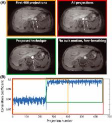
          Non‐Cartesian magnetic resonance imaging (MRI) sequences have shown great promise for abdominal examination during free breathing, but break down in the presence of bulk patient motion (i.e. voluntary or involuntary patient movement resulting in translation, rotation or elastic deformations of the body). This work describes a data‐consistency‐driven image stabilization technique that detects and excludes bulk movements during data acquisition. Bulk motion is identified from changes in the signal intensity distribution across different elements of a multi‐channel receive coil array. A short free induction decay signal is acquired after excitation and used as a measure to determine alterations in the load distribution. The technique has been implemented on a clinical MR scanner and evaluated in the abdomen. Six volunteers were scanned and two radiologists scored the reconstructions. To show the applicability to other body areas, additional neck and knee images were acquired. Data corrupted by bulk motion were successfully detected and excluded from image reconstruction. An overall increase in image sharpness and reduction of streaking and shine‐through artifacts were seen in the volunteer study, as well as in the neck and knee scans. The proposed technique enables automatic real‐time detection and exclusion of bulk motion during MR examinations without user interaction. It may help to improve the reliability of pediatric MRI examinations without the use of sedation.

          Related collections

          Most cited references31

          • Record: found
          • Abstract: found
          • Article: not found

          An optimal radial profile order based on the Golden Ratio for time-resolved MRI.

          In dynamic magnetic resonance imaging (MRI) studies, the motion kinetics or the contrast variability are often hard to predict, hampering an appropriate choice of the image update rate or the temporal resolution. A constant azimuthal profile spacing (111.246 degrees), based on the Golden Ratio, is investigated as optimal for image reconstruction from an arbitrary number of profiles in radial MRI. The profile order is evaluated and compared with a uniform profile distribution in terms of signal-to-noise ratio (SNR) and artifact level. The favorable characteristics of such a profile order are exemplified in two applications on healthy volunteers. First, an advanced sliding window reconstruction scheme is applied to dynamic cardiac imaging, with a reconstruction window that can be flexibly adjusted according to the extent of cardiac motion that is acceptable. Second, a contrast-enhancing k-space filter is presented that permits reconstructing an arbitrary number of images at arbitrary time points from one raw data set. The filter was utilized to depict the T1-relaxation in the brain after a single inversion prepulse. While a uniform profile distribution with a constant angle increment is optimal for a fixed and predetermined number of profiles, a profile distribution based on the Golden Ratio proved to be an appropriate solution for an arbitrary number of profiles.
            Bookmark
            • Record: found
            • Abstract: found
            • Article: not found

            Motion artifacts in MRI: A complex problem with many partial solutions.

            Subject motion during magnetic resonance imaging (MRI) has been problematic since its introduction as a clinical imaging modality. While sensitivity to particle motion or blood flow can be used to provide useful image contrast, bulk motion presents a considerable problem in the majority of clinical applications. It is one of the most frequent sources of artifacts. Over 30 years of research have produced numerous methods to mitigate or correct for motion artifacts, but no single method can be applied in all imaging situations. Instead, a "toolbox" of methods exists, where each tool is suitable for some tasks, but not for others. This article reviews the origins of motion artifacts and presents current mitigation and correction methods. In some imaging situations, the currently available motion correction tools are highly effective; in other cases, appropriate tools still need to be developed. It seems likely that this multifaceted approach will be what eventually solves the motion sensitivity problem in MRI, rather than a single solution that is effective in all situations. This review places a strong emphasis on explaining the physics behind the occurrence of such artifacts, with the aim of aiding artifact detection and mitigation in particular clinical situations.
              Bookmark
              • Record: found
              • Abstract: found
              • Article: not found

              Prospective motion correction in brain imaging: a review.

              Motion correction in magnetic resonance imaging by real-time adjustment of the imaging pulse sequence was first proposed more than 20 years ago. Recent advances have resulted from combining real-time correction with new navigator and external tracking mechanisms capable of quantifying rigid-body motion in all 6 degrees of freedom. The technique is now often referred to as "prospective motion correction." This article describes the fundamentals of prospective motion correction and reviews the latest developments in its application to brain imaging and spectroscopy. Although emphasis is placed on the brain as the organ of interest, the same principles apply whenever the imaged object can be approximated as a rigid body. Prospective motion correction can be used with most MR sequences, so it has potential to make a large impact in clinical routine. To maximize the benefits obtained from the technique, there are, however, several challenges still to be met. These include practical implementation issues, such as obtaining tracking data with minimal delay, and more fundamental problems, such as the magnetic field distortions caused by a moving object. This review discusses these challenges and summarizes the state of the art. We hope that this work will motivate further developments in prospective motion correction and help the technique to reach its full potential. Copyright © 2012 Wiley Periodicals, Inc.
                Bookmark

                Author and article information

                Contributors
                b.stemkens@umcutrecht.nl
                Journal
                NMR Biomed
                NMR Biomed
                10.1002/(ISSN)1099-1492
                NBM
                Nmr in Biomedicine
                John Wiley and Sons Inc. (Hoboken )
                0952-3480
                1099-1492
                08 September 2017
                November 2017
                : 30
                : 11 ( doiID: 10.1002/nbm.v30.11 )
                : e3830
                Affiliations
                [ 1 ] Department of Radiotherapy University Medical Center Utrecht the Netherlands
                [ 2 ] Center for Advanced Imaging Innovation and Research (CAI 2R), Department of Radiology New York University School of Medicine New York NY USA
                [ 3 ] Bernard and Irene Schwartz Center for Biomedical Imaging, Department of Radiology New York University School of Medicine New York NY USA
                Author notes
                [*] [* ] Correspondence

                B. Stemkens, Department of Radiotherapy, UMC Utrecht, Heidelberglaan 100, 3584CX Utrecht, the Netherlands.

                Email: b.stemkens@ 123456umcutrecht.nl

                Author information
                http://orcid.org/0000-0003-0488-8070
                Article
                NBM3830 NBM-17-0178.R1
                10.1002/nbm.3830
                5643254
                28885742
                8fb024d0-e998-41e8-a429-ec2fa1d720ce
                © 2017 The Authors. NMR in Biomedicine published by John Wiley & Sons Ltd.

                This is an open access article under the terms of the Creative Commons Attribution License, which permits use, distribution and reproduction in any medium, provided the original work is properly cited.

                History
                : 18 July 2017
                : 03 August 2017
                : 14 August 2017
                Page count
                Figures: 6, Tables: 2, Pages: 11, Words: 5689
                Funding
                Funded by: US National Institute of Health
                Award ID: NIH P41 EB017183
                Award ID: NIH R01 EB018308
                Funded by: Royal Netherlands Academy of Arts and Sciences (KNAW) Ter Meulen grant
                Categories
                Research Article
                Research Articles
                Custom metadata
                2.0
                nbm3830
                November 2017
                Converter:WILEY_ML3GV2_TO_NLMPMC version:5.2.1 mode:remove_FC converted:26.10.2017

                Radiology & Imaging
                abdominal imaging,bulk motion,motion correction,prospective motion detection,real‐time motion detection

                Comments

                Comment on this article