20
views
0
recommends
+1 Recommend
0 collections
    0
    shares
      • Record: found
      • Abstract: found
      • Article: found
      Is Open Access

      Accuracy of abdominal ultrasound for the diagnosis of pneumoperitoneum in patients with acute abdominal pain: a pilot study

      research-article

      Read this article at

      Bookmark
          There is no author summary for this article yet. Authors can add summaries to their articles on ScienceOpen to make them more accessible to a non-specialist audience.

          Abstract

          Background

          Pneumoperitoneum is a rare cause of abdominal pain characterized by a high mortality. Ultrasonography (US) can detect free intraperitoneal air; however, its accuracy remains unclear. The aims of this pilot study were to define the diagnostic performance and the reliability of abdominal US for the diagnosis of pneumoperitoneum.

          Methods

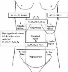
          This was a prospective observational study. Four senior and two junior physicians were shown, in an unpaired randomized order, abdominal US videos from 11 patients with and 11 patients without pneumoperitoneum. Abdominal US videos were obtained from consecutive patients presenting to ED complaining abdominal pain with the diagnosis of pneumoperitoneum established by CT. Abdominal US was performed according to a standardized protocol that included the following scans: epigastrium, right and left hypochondrium, umbilical area and right hypochondrium with the patient lying on the left flank. We evaluated accuracy, intra- and inter-observer agreement of abdominal US when reviewed by senior physicians. Furthermore, we compared the accuracy of a “2 scan-fast exam” (epigastrium and right hypochondrium) vs the full US examination and the accuracy of physicians expert in US vs nonexpert ones. Finally, accuracy of US was compared with abdominal radiography in patients with available images.

          Results

          Considering senior revision, accuracy of abdominal US was 88.6 % (95 % CI 79.4-92.4 %) with a sensitivity of 95.5 % (95 % CI 86.3–99.2 %) and a specificity of 81.8 % (95 % CI 72.6–85.5 %). Inter- and intra-observer agreement (k) were 0.64 and 0.95, respectively. Accuracy of a “2 scan-fast exam” (87.5 %, 95 % CI 77.9–92.4 %) was similar to global exam. Sensitivity of abdominal radiography (72.2 %, 95 % CI 54.8–85.7 %) was lower than that of abdominal US, while specificity (92.5 %, 95 % CI 79.5–98.3 %) was higher. Accuracy (68.2 %, 95 % CI 51.4–80.9 %) of junior reviewers evaluating US was lower than senior reviewers.

          Conclusions

          Senior physicians can recognize US signs of pneumoperitoneum with a good accuracy and reliability; sensitivity of US could be superior to abdominal radiography and a 2 fast-scan exam seems as accurate as full abdominal examination. US could be a useful bedside screening test for pneumoperitoneum.

          Trial registry ClinicalTrials.gov; No.: NCT02004925; URL: http://www.clinicaltrials.gov

          Electronic supplementary material

          The online version of this article (doi:10.1186/s13089-015-0032-6) contains supplementary material, which is available to authorized users.

          Related collections

          Most cited references14

          • Record: found
          • Abstract: found
          • Article: not found

          Selective use of ultrasonography for the detection of pneumoperitoneum.

          Whether ultrasonography is superior to plain radiography for the detection of pneumoperitoneum is unknown. The goal of this study was to determine the value of ultrasonography for the detection of pneumoperitoneum. One hundred thirty-two patients with suspected hollow-organ perforation were prospectively selected for study. All 132 patients received ultrasonography, upright chest radiography, and left lateral decubitus abdominal radiography examinations. The diagnostic accuracies of chest and abdominal radiographs for the detection of pneumoperitoneum were compared with corresponding values from ultrasonography. Of the 125 patients who underwent laparotomy, 121 patients had hollow-organ perforation, three patients had perforated appendicitis, and one patient had acute cholecystitis. For the diagnosis of pneumoperitoneum, ultrasonography demonstrated a sensitivity of 93%, a specificity of 64%, a positive predictive value of 97%, a negative predictive value of 44%, and an accuracy of 90%. Plain radiography revealed a sensitivity of 79%, a specificity of 64%, a positive predictive value of 96%, a negative predictive value of 21%, and an accuracy of 77%. Ultrasonography is a more sensitive diagnosing modality than plain radiography for the diagnosis of pneumoperitoneum. The authors suggest that ultrasonography was a useful diagnostic modality when plain radiographs failed to reveal pneumoperitoneum among patients with suspected hollow-organ perforation.
            Bookmark
            • Record: found
            • Abstract: found
            • Article: not found

            Ultrasonography is superior to plain radiography in the diagnosis of pneumoperitoneum.

            The aim of this study was to compare plain radiography with abdominal ultrasonography in the detection of pneumoperitoneum. A total of 188 patients with suspected hollow organ perforation were studied. All patients had abdominal ultrasonography, upright chest radiography and left lateral decubitus abdominal radiography examinations. The sensitivity, specificity, positive and negative predictive value, and accuracy of chest and abdominal radiography were compared with that of abdominal ultrasonography. One hundred and seventy-eight patients underwent laparotomy; 170 patients had hollow organ perforation, five patients had perforated appendicitis and three had acute cholecystitis. In the diagnosis of pneumoperitoneum, ultrasonography had improved sensitivity (92 versus 78 per cent), negative predictive value (39 versus 20 per cent) and accuracy (88 versus 76 per cent), and similar specificity (both 53 per cent) and positive predictive value (95 versus 94 per cent) compared with plain radiography. Ultrasonography is more sensitive than plain radiography in the diagnosis of pneumoperitoneum.
              Bookmark
              • Record: found
              • Abstract: found
              • Article: not found

              Focus on abnormal air: diagnostic ultrasonography for the acute abdomen.

              Emergency ultrasonography is a frequently used imaging tool in the bedside diagnosis of the acute abdomen. Classic indications include imaging for acute abdominal aneurysm, acute cholecystitis, hydronephrosis, and free intra-abdominal fluid in patients with trauma or suspected vascular or ectopic pregnancy rupture. Point-of-care sonographic imaging often emphasizes the diagnostic utility of fluid and edema, both as a significant finding and as a desirable adjunct for improved imaging. Conversely, the finding of sonographic intra-abdominal air is commonly 'tolerated' as a necessary evil that can foil image acquisition. This is in stark contrast to the accepted diagnostic utility of air in other imaging modalities for the acute abdomen, such as computed tomography and conventional radiography. Countering the bias against air as a deterrent for diagnostic ultrasound's accuracy are several published studies suggesting that abnormal air patterns can be used with high precision to diagnose pneumoperitoneum. These studies advocate that sonographic findings of abnormal air can be straightforward and can become crucial for increasing the diagnostic yield of bedside ultrasound of the acute abdomen. They suggest that practitioners should familiarize themselves with the findings and techniques to gain the experience required to make the diagnosis with confidence. This article will discuss four groups of abnormal air patterns found in the abdomen and the retroperitoneum and the respective scanning techniques, with a focus on the use of ultrasound for diagnosing pneumoperitoneum and a suggested scanning approach in the emergency setting.
                Bookmark

                Author and article information

                Contributors
                0039 3396122448 , pnazerian@hotmail.com
                Journal
                Crit Ultrasound J
                Crit Ultrasound J
                Critical Ultrasound Journal
                Springer Milan (Milan )
                2036-3176
                2036-7902
                6 October 2015
                6 October 2015
                2015
                : 7
                : 15
                Affiliations
                [ ]Department of Emergency Medicine, Careggi University Hospital, largo Brambilla 3, 50134 Florence, Italy
                [ ]Radiology Department, Careggi University Hospital, Florence, Italy
                [ ]Department of Emergency Medicine, Pinerolo Hospital, Turin, Italy
                Article
                32
                10.1186/s13089-015-0032-6
                4595408
                26443344
                8fcb43c8-1ce0-48cb-a5cf-308fdb6b56f5
                © Nazerian et al. 2015

                Open AccessThis article is distributed under the terms of the Creative Commons Attribution 4.0 International License ( http://creativecommons.org/licenses/by/4.0/), which permits unrestricted use, distribution, and reproduction in any medium, provided you give appropriate credit to the original author(s) and the source, provide a link to the Creative Commons license, and indicate if changes were made.

                History
                : 15 May 2015
                : 16 September 2015
                Categories
                Original Article
                Custom metadata
                © The Author(s) 2015

                Radiology & Imaging
                pneumoperitoneum diagnosis,abdominal ultrasound,abdominal radiography,abdominal pain,hollow organ perforation diagnosis

                Comments

                Comment on this article