12
views
0
recommends
+1 Recommend
0 collections
    0
    shares
      • Record: found
      • Abstract: found
      • Article: found
      Is Open Access

      Large Extracellular Vesicle Characterization and Association with Circulating Tumor Cells in Metastatic Castrate Resistant Prostate Cancer

      research-article

      Read this article at

      Bookmark
          There is no author summary for this article yet. Authors can add summaries to their articles on ScienceOpen to make them more accessible to a non-specialist audience.

          Abstract

          Simple Summary

          Non-invasive, liquid biopsies are an attractive means for tumor diagnosis and monitoring. In addition to DNA and cells, tumors have an increased propensity compared to normal cells to shed vesicles. These large extracellular vesicles, which are believed to be more frequent in aggressive cancers, carry tumor DNA and proteins and can thus be informative sources for diagnosis and prognosis. In this study, we developed a method to identify and molecularly characterize large extracellular vesicles in parallel to circulating tumor cells at a single-cell/single-vesicle level. We show that the number of large extracellular vesicles correlates to and exceeds the number of circulating tumor cells, and that analysis of tumor-derived large extracellular vesicles hence increases the sensitivity of the liquid biopsy assay.

          Abstract

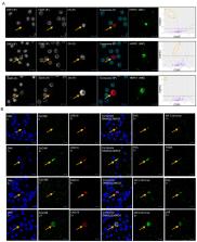
          Liquid biopsies hold potential as minimally invasive sources of tumor biomarkers for diagnosis, prognosis, therapy prediction or disease monitoring. We present an approach for parallel single-object identification of circulating tumor cells (CTCs) and tumor-derived large extracellular vesicles (LEVs) based on automated high-resolution immunofluorescence followed by downstream multiplexed protein profiling. Identification of LEVs >6 µm in size and CTC enumeration was highly correlated, with LEVs being 1.9 times as frequent as CTCs, and additional LEVs were identified in 73% of CTC-negative liquid biopsy samples from metastatic castrate resistant prostate cancer. Imaging mass cytometry (IMC) revealed that 49% of cytokeratin (CK)-positive LEVs and CTCs were EpCAM-negative, while frequently carrying prostate cancer tumor markers including AR, PSA, and PSMA. HSPD1 was shown to be a specific biomarker for tumor derived circulating cells and LEVs. CTCs and LEVs could be discriminated based on size, morphology, DNA load and protein score but not by protein signatures. Protein profiles were overall heterogeneous, and clusters could be identified across object classes. Parallel analysis of CTCs and LEVs confers increased sensitivity for liquid biopsies and expanded specificity with downstream characterization. Combined, it raises the possibility of a more comprehensive assessment of the disease state for precise diagnosis and monitoring.

          Related collections

          Most cited references43

          • Record: found
          • Abstract: found
          • Article: not found

          Tumour exosome integrins determine organotropic metastasis

          Ever since Stephen Paget’s 1889 hypothesis, metastatic organotropism has remained one of cancer’s greatest mysteries. Here we demonstrate that exosomes from mouse and human lung-, liver- and brain-tropic tumour cells fuse preferentially with resident cells at their predicted destination, namely lung fibroblasts and epithelial cells, liver Kupffer cells and brain endothelial cells. We show that tumour-derived exosomes uptaken by organ-specific cells prepare the pre-metastatic niche. Treatment with exosomes from lung-tropic models redirected the metastasis of bone-tropic tumour cells. Exosome proteomics revealed distinct integrin expression patterns, in which the exosomal integrins α6β4 and α6β1 were associated with lung metastasis, while exosomal integrin αvβ5 was linked to liver metastasis. Targeting the integrins α6β4 and αvβ5 decreased exosome uptake, as well as lung and liver metastasis, respectively. We demonstrate that exosome integrin uptake by resident cells activates Src phosphorylation and pro-inflammatory S100 gene expression. Finally, our clinical data indicate that exosomal integrins could be used to predict organ-specific metastasis.
            Bookmark
            • Record: found
            • Abstract: found
            • Article: not found

            Proteomic comparison defines novel markers to characterize heterogeneous populations of extracellular vesicle subtypes.

            Extracellular vesicles (EVs) have become the focus of rising interest because of their numerous functions in physiology and pathology. Cells release heterogeneous vesicles of different sizes and intracellular origins, including small EVs formed inside endosomal compartments (i.e., exosomes) and EVs of various sizes budding from the plasma membrane. Specific markers for the analysis and isolation of different EV populations are missing, imposing important limitations to understanding EV functions. Here, EVs from human dendritic cells were first separated by their sedimentation speed, and then either by their behavior upon upward floatation into iodixanol gradients or by immuno-isolation. Extensive quantitative proteomic analysis allowing comparison of the isolated populations showed that several classically used exosome markers, like major histocompatibility complex, flotillin, and heat-shock 70-kDa proteins, are similarly present in all EVs. We identified proteins specifically enriched in small EVs, and define a set of five protein categories displaying different relative abundance in distinct EV populations. We demonstrate the presence of exosomal and nonexosomal subpopulations within small EVs, and propose their differential separation by immuno-isolation using either CD63, CD81, or CD9. Our work thus provides guidelines to define subtypes of EVs for future functional studies.
              Bookmark
              • Record: found
              • Abstract: found
              • Article: not found

              Extracellular vesicles in cancer — implications for future improvements in cancer care

              The sustained growth, invasion, and metastasis of cancer cells depend upon bidirectional cell-cell communication within complex tissue environments. Such communication predominantly involves the secretion of soluble factors by cancer cells and/or stromal cells within the tumour microenvironment (TME), although these cell types have also been shown to export membrane-encapsulated particles containing regulatory molecules that contribute to cell-cell communication. These particles are known as extracellular vesicles (EVs) and include species of exosomes and shed microvesicles. EVs carry molecules such as oncoproteins and oncopeptides, RNA species (for example, microRNAs, mRNAs, and long non-coding RNAs), lipids, and DNA fragments from donor to recipient cells, initiating profound phenotypic changes in the TME. Emerging evidence suggests that EVs have crucial roles in cancer development, including pre-metastatic niche formation and metastasis. Cancer cells are now recognized to secrete more EVs than their nonmalignant counterparts, and these particles can be isolated from bodily fluids. Thus, EVs have strong potential as blood-based or urine-based biomarkers for the diagnosis, prognostication, and surveillance of cancer. In this Review, we discuss the biophysical properties and physiological functions of EVs, particularly their pro-metastatic effects, and highlight the utility of EVs for the development of cancer diagnostics and therapeutics.
                Bookmark

                Author and article information

                Contributors
                Role: Academic Editor
                Role: Academic Editor
                Journal
                Cancers (Basel)
                Cancers (Basel)
                cancers
                Cancers
                MDPI
                2072-6694
                02 March 2021
                March 2021
                : 13
                : 5
                : 1056
                Affiliations
                [1 ]USC Michelson Center for Convergent Bioscience, University of Southern California, Los Angeles, CA 90089, USA; anna.sandstrom_gerdtsson@ 123456immun.lth.se (A.S.G.); msetayes@ 123456usc.edu (S.M.S.); Peymaneh87@ 123456gmail.com (P.D.M.); ruizvela@ 123456usc.edu (C.R.); anders@ 123456bionamic.io (A.C.); rnevarez@ 123456usc.edu (R.N.); matsumon@ 123456usc.edu (N.M.); erik.gerdtsson@ 123456gmail.com (E.G.); jameshic@ 123456usc.edu (J.H.)
                [2 ]Department of Genitourinary Medical Oncology, The University of Texas MD Anderson Cancer Center, Houston, TX 77030, USA; azurita@ 123456mdanderson.org (A.Z.); clogothe@ 123456mdanderson.org (C.L.); PCorn@ 123456mdanderson.org (P.G.C.); aaparicio@ 123456mdanderson.org (A.M.A.)
                Author notes
                [* ]Correspondence: pkuhn@ 123456usc.edu ; Tel.: +1-213-821-3980
                Author information
                https://orcid.org/0000-0003-1932-0365
                Article
                cancers-13-01056
                10.3390/cancers13051056
                7958848
                33801459
                902db16f-cb61-49a9-8949-1318c993c1f2
                © 2021 by the authors.

                Licensee MDPI, Basel, Switzerland. This article is an open access article distributed under the terms and conditions of the Creative Commons Attribution (CC BY) license ( http://creativecommons.org/licenses/by/4.0/).

                History
                : 20 January 2021
                : 24 February 2021
                Categories
                Article

                large extracellular vesicles,oncosomes,ctcs,liquid biopsy,metastatic castrate-resistant prostate cancer,aggressive variant prostate cancer

                Comments

                Comment on this article