7
views
0
recommends
+1 Recommend
0 collections
    0
    shares
      • Record: found
      • Abstract: found
      • Article: found
      Is Open Access

      Heads-Up 3D Surgery under Low Light Intensity Conditions: New High-Sensitivity HD Camera for Ophthalmological Microscopes

      research-article

      Read this article at

      Bookmark
          There is no author summary for this article yet. Authors can add summaries to their articles on ScienceOpen to make them more accessible to a non-specialist audience.

          Abstract

          Purpose

          To determine the feasibility of performing intraocular surgeries in a heads-up position with low illuminance conditions by observing a display of the surgical field created by a three-dimensional imaging (3D) system.

          Methods

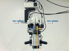
          Seventy-four eyes of 56 patients underwent cataract surgery (72 eyes) with the heads-up 3D surgery system; 60 eyes with cataract surgery alone, 7 eyes with combined cataract and glaucoma microdevice implant surgery, 5 eyes with combined cataract and vitrectomy surgery, and two eyes with vitrectomy surgery alone were studied. The illuminance from the surgical microscope was set to be dimmer (Leica M822F40 main light 2%; otto-flex 6%) than the usual setting to minimize the discomfort and glare for the patient. The surgeries were performed under topical anesthesia. The luminance of the images observed through the eyepieces of the operating microscope and the image of a 3D system created by a high-sensitivity sensor Exmor R 3CMOS HD camera (Sony MCC-1000MD) were measured.

          Results

          All surgeries were completed without any complications under the low illumination conditions. The surgical field on the display monitor was created by a 3D system using a high-sensitivity sensor camera and was observed in a heads-up position. The patients did not report any intolerable discomfort or glare during the surgery. Cataract surgeries were performed with a good view of the surgical field under the extremely low illumination from the surgical microscope. The high-sensitivity sensors and electronic amplifications of the image signals made the surgical field brighter and allowed the surgeon to perform the surgery confidently and safely.

          Conclusions

          Heads-up, 3D-assisted intraocular surgeries can be performed safely and efficiently with low illuminance of the surgical field. This trial is registered with UMIN000037838.

          Related collections

          Most cited references8

          • Record: found
          • Abstract: found
          • Article: not found

          HEADS-UP SURGERY FOR VITREORETINAL PROCEDURES: An Experimental and Clinical Study.

          To investigate the feasibility of performing vitrectomies while viewing a three-dimensional image on a large display in a heads-up position.
            Bookmark
            • Record: found
            • Abstract: found
            • Article: not found

            MINIMAL ENDOILLUMINATION LEVELS AND DISPLAY LUMINOUS EMITTANCE DURING THREE-DIMENSIONAL HEADS-UP VITREORETINAL SURGERY.

            To determine minimal endoillumination levels required to perform 3-dimensional heads-up vitreoretinal surgery and to correlate endoillumination levels used for measurements of heads-up display (HUD) luminous emittance.
              Bookmark
              • Record: found
              • Abstract: found
              • Article: found
              Is Open Access

              Comparison of clinical outcomes between “heads-up” 3D viewing system and conventional microscope in macular hole surgeries: A pilot study

              Purpose: To compare clinical outcomes of patients undergoing macular hole surgery with heads-up three-dimensional (3D) viewing system and conventional microscope. Methods: In all, 50 eyes of 50 patients with stage 3 or 4 macular hole were randomized and macular hole surgery [inverted internal limiting membrane (ILM) flap technique] was performed in 25 eyes using 3D viewing system and 25 eyes using conventional microscope. All surgeries were performed by a single surgeon. Patients were followed up for a period of 3 months. Logarithm of the minimum angle of resolution (logMAR) visual acuity, macular hole index, intraoperative parameters such as total surgical time, total ILM peel time, number of flap initiations, duration of Brilliant Blue G dye exposure, illumination intensity, postoperative logMAR visual acuity, and macular hole closure rates were recorded and compared between the two groups. Results: The mean age was 67.92 ± 7.95 and 67.96 ± 4.78 years in both groups, respectively (P = 0.98). Gender (P = 0.38) and right versus left eye (P = 0.39) were also comparable. Preoperative and postoperative best-corrected visual acuity (P = 0.86, 0.92), macular hole index (P = 0.96), total surgical time (P = 0.56), total ILM peel time (P = 0.49), number of flap initiations (P = 0.11), and macular hole closure rates (P = 0.61) were not statistically significant when compared between the two groups. Illumination intensity of microscope (100% vs 45%) and endoillumination (40% vs 13%) were significantly less in the 3D viewing system. Conclusion: The clinical outcomes of macular hole surgery using 3D viewing system are not inferior to that of conventional microscopes, and it has the added advantages of better ergonomics, reduced phototoxicity, peripheral visualization, magnification, and less asthenopia, and it serves as a good educational tool.
                Bookmark

                Author and article information

                Contributors
                Journal
                J Ophthalmol
                J Ophthalmol
                JOPH
                Journal of Ophthalmology
                Hindawi
                2090-004X
                2090-0058
                2019
                25 November 2019
                : 2019
                : 5013463
                Affiliations
                1Department of Ophthalmology, Teikyo University School of Medicine, Kaga 2-11-1, Itabashi-ku, Tokyo 173-8605, Japan
                2Department of Ophthalmology, Saitama Medical University School of Medicine, Morohongo 38, Moroyama-machi, Saitama 350-0495, Japan
                3Matsumoto Eye Clinic, Tokushima, Japan
                4Ohno Eye Clinic, Saitama, Japan
                Author notes

                Academic Editor: Masaru Takeuchi

                Author information
                https://orcid.org/0000-0003-0227-1106
                https://orcid.org/0000-0002-5464-2573
                https://orcid.org/0000-0002-1543-9345
                Article
                10.1155/2019/5013463
                6899266
                31885885
                90a020b4-570b-4d3e-9ebc-4b47eb1616f0
                Copyright © 2019 Celso Soiti Matsumoto et al.

                This is an open access article distributed under the Creative Commons Attribution License, which permits unrestricted use, distribution, and reproduction in any medium, provided the original work is properly cited.

                History
                : 4 August 2019
                : 7 November 2019
                Funding
                Funded by: Matsumoto Eye Clinic
                Categories
                Clinical Study

                Ophthalmology & Optometry
                Ophthalmology & Optometry

                Comments

                Comment on this article