31
views
0
recommends
+1 Recommend
0 collections
    0
    shares
      • Record: found
      • Abstract: found
      • Article: found
      Is Open Access

      Structural Insights for Activation of Retinal Guanylate Cyclase by GCAP1

      research-article

      Read this article at

      Bookmark
          There is no author summary for this article yet. Authors can add summaries to their articles on ScienceOpen to make them more accessible to a non-specialist audience.

          Abstract

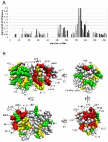
          Guanylyl cyclase activating protein 1 (GCAP1), a member of the neuronal calcium sensor (NCS) subclass of the calmodulin superfamily, confers Ca 2+-sensitive activation of retinal guanylyl cyclase 1 (RetGC1) upon light activation of photoreceptor cells. Here we present NMR assignments and functional analysis to probe Ca 2+-dependent structural changes in GCAP1 that control activation of RetGC. NMR assignments were obtained for both the Ca 2+-saturated inhibitory state of GCAP1 versus a GCAP1 mutant (D144N/D148G, called EF4mut), which lacks Ca 2+ binding in EF-hand 4 and models the Ca 2+-free/Mg 2+-bound activator state of GCAP1. NMR chemical shifts of backbone resonances for Ca 2+-saturated wild type GCAP1 are overall similar to those of EF4mut, suggesting a similar main chain structure for assigned residues in both the Ca 2+-free activator and Ca 2+-bound inhibitor states. This contrasts with large Ca 2+-induced chemical shift differences and hence dramatic structural changes seen for other NCS proteins including recoverin and NCS-1. The largest chemical shift differences between GCAP1 and EF4mut are seen for residues in EF4 (S141, K142, V145, N146, G147, G149, E150, L153, E154, M157, E158, Q161, L166), but mutagenesis of EF4 residues (F140A, K142D, L153R, L166R) had little effect on RetGC1 activation. A few GCAP1 residues in EF-hand 1 (K23, T27, G32) also show large chemical shift differences, and two of the mutations (K23D and G32N) each decrease the activation of RetGC, consistent with a functional conformational change in EF1. GCAP1 residues at the domain interface (V77, A78, L82) have NMR resonances that are exchange broadened, suggesting these residues may be conformationally dynamic, consistent with previous studies showing these residues are in a region essential for activating RetGC1.

          Related collections

          Most cited references62

          • Record: found
          • Abstract: found
          • Article: not found

          Neuronal calcium sensor proteins: generating diversity in neuronal Ca2+ signalling.

          In neurons, intracellular calcium signals have crucial roles in activating neurotransmitter release and in triggering alterations in neuronal function. Calmodulin has been widely studied as a Ca(2+) sensor that has several defined roles in neuronal Ca(2+) signalling, but members of the neuronal calcium sensor protein family have also begun to emerge as key components in a number of regulatory pathways and have increased the diversity of neuronal Ca(2+) signalling pathways. The differing properties of these proteins allow them to have discrete, non-redundant functions.
            Bookmark
            • Record: found
            • Abstract: found
            • Article: not found

            Backbone dynamics of Escherichia coli ribonuclease HI: correlations with structure and function in an active enzyme.

            Ribonuclease H is an endonuclease that hydrolyzes the RNA moiety of RNA-DNA duplex molecules. Escherichia coli ribonuclease H is involved in DNA replication, and retroviral ribonuclease H is essential for reverse transcription of the viral genome. To characterize the intramolecular dynamical properties of E. coli ribonuclease H, spin-lattice relaxation rate constants, spin-spin relaxation rate constants and steady state nuclear Overhauser effects for the 15N nuclear spins were measured by using proton-detected heteronuclear NMR spectroscopy. The relaxation data were analyzed by using a series of dynamical models in conjunction with a statistical model selection protocol. Ribonuclease H exhibits a complex array of dynamical features, most notably in the parallel beta-strands of the principal five-stranded beta-sheet, the coiled-coil helical interface, the active site, and the loop regions surrounding the active site. The dynamical properties are correlated with local structural environments of the 15N spins and suggest possible relationships to the functional properties of ribonuclease H. Results for E. coli ribonuclease H are compared to previously reported results for the human immunodeficiency virus type 1 ribonuclease H domain of reverse transcriptase.
              Bookmark
              • Record: found
              • Abstract: found
              • Article: not found

              The neuronal calcium sensor family of Ca2+-binding proteins.

              Ca(2+) plays a central role in the function of neurons as the trigger for neurotransmitter release, and many aspects of neuronal activity, from rapid modulation to changes in gene expression, are controlled by Ca(2+). These actions of Ca(2+) must be mediated by Ca(2+)-binding proteins, including calmodulin, which is involved in Ca(2+) regulation, not only in neurons, but in most other cell types. A large number of other EF-hand-containing Ca(2+)-binding proteins are known. One family of these, the neuronal calcium sensor (NCS) proteins, has a restricted expression in retinal photoreceptors or neurons and neuroendocrine cells, suggesting that they have specialized roles in these cell types. Two members of the family (recoverin and guanylate cyclase-activating protein) have established roles in the regulation of phototransduction. Despite close sequence similarities, the NCS proteins have distinct neuronal distributions, suggesting that they have different functions. Recent work has begun to demonstrate the physiological roles of members of this protein family. These include roles in the modulation of neurotransmitter release, control of cyclic nucleotide metabolism, biosynthesis of polyphosphoinositides, regulation of gene expression and in the direct regulation of ion channels. In the present review we describe the known sequences and structures of the NCS proteins, information on their interactions with target proteins and current knowledge about their cellular and physiological functions.
                Bookmark

                Author and article information

                Contributors
                Role: Editor
                Journal
                PLoS One
                PLoS ONE
                plos
                plosone
                PLoS ONE
                Public Library of Science (San Francisco, USA )
                1932-6203
                2013
                13 November 2013
                : 8
                : 11
                : e81822
                Affiliations
                [1 ]Department of Chemistry, University of California Davis, Davis, California, United States of America
                [2 ]Basic Sciences, Pennsylvania College of Optometry, Salus University, Elkins Park, Pennsylvania, United States of America
                University of Oldenburg, Germany
                Author notes

                Competing Interests: The authors have declared that no competing interests exist.

                Conceived and designed the experiments: JBA AMD. Performed the experiments: SL IVP. Analyzed the data: SL JBA. Contributed reagents/materials/analysis tools: JBA AMD. Wrote the manuscript: JBA AMD.

                Article
                PONE-D-13-35164
                10.1371/journal.pone.0081822
                3827477
                24236217
                92d3b796-8b38-4ebb-ab77-f8ebff95b35b
                Copyright @ 2013

                This is an open-access article distributed under the terms of the Creative Commons Attribution License, which permits unrestricted use, distribution, and reproduction in any medium, provided the original author and source are credited.

                History
                : 26 August 2013
                : 27 October 2013
                Funding
                This work was funded by NIH grants EY012347 (J.B.A.), EY11522 (A.M.D.) and RR11973 (UC Davis NMR). The URL for NIH is www.nih.gov. The funders had no role in study design, data collection and analysis, decision to publish, or preparation of the manuscript.
                Categories
                Research Article

                Uncategorized
                Uncategorized

                Comments

                Comment on this article