0
views
0
recommends
+1 Recommend
0 collections
    0
    shares
      • Record: found
      • Abstract: found
      • Article: found
      Is Open Access

      Sonographic assessment of the tarsal tunnel compared to cadaveric findings: a pictorial study

      research-article
      1 , 2 , 3 , 2 ,
      Journal of Ultrasonography
      Sciendo
      ultrasound, anatomy, tarsal tunnel

      Read this article at

      Bookmark
          There is no author summary for this article yet. Authors can add summaries to their articles on ScienceOpen to make them more accessible to a non-specialist audience.

          Abstract

          Aim of the study

          To present the anatomy of the tarsal tunnel and demonstrate the utility of high-resolution ultrasound for tarsal tunnel examination.

          Materials and methods

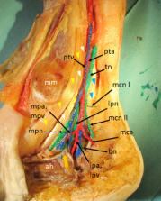
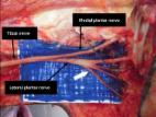
          Anatomical dissection was performed on a defrosted cadaveric model to demonstrate relevant anatomical structures of the tarsal tunnel, namely tendons, vessels and nerves. The tibial nerve division was demonstrated; the bifurcation of the tibial nerve into the medial and lateral plantar nerve, two medial calcaneal nerve branches were identified originating from the tibial nerve and the Baxter’s nerve was identified as the first branch of the lateral plantar nerve. An ultrasound examination of the tarsal tunnel region was performed on a healthy volunteer. A linear probe was used and sonographic images were obtained at different levels of the tarsal tunnel: the proximal tarsal tunnel, the tibial nerve division into the medial and lateral plantar nerves, the distal tarsal tunnel, the Baxter’s nerve branching point and the Baxter’s nerve crossing between the abductor hallucis and quadratus plantae muscle.

          Results

          Sonographic images were correlated with anatomical structures exposed during cadaveric dissection.

          Conclusions

          We presented the anatomic-sonographic correlation of the tarsal tunnel and showed that high-resolution ultrasound is a useful imaging modality for tarsal tunnel assessment.

          Related collections

          Most cited references21

          • Record: found
          • Abstract: found
          • Article: not found

          Tibial nerve branching in the tarsal tunnel.

          To provide an anatomical basis for diagnosis and treatment of the tarsal tunnel syndrome, the relationship of the tibial nerve to the tarsal tunnel was investigated in 31 feet of 20 cadavers. The bifurcation into medial and lateral plantar nerves occurred within 1 cm of the malleolar-calcaneal axis in 90% of the feet. Seven of 11 bilateral specimens were bilaterally symmetrical in the bifurcation location; three varied within 1 cm between sides; and in the fourth cadaver, one side bifurcated at 3 cm and the other at 5 cm proximal to the axis. The calcaneal nerve showed great variability; in seven cadavers, it arose within, in eight cadavers proximal to, and in five cadavers there were multiple branches arising both proximal to and within the tarsal tunnel.
            Bookmark
            • Record: found
            • Abstract: found
            • Article: found
            Is Open Access

            Clinical-anatomic mapping of the tarsal tunnel with regard to Baxter’s neuropathy in recalcitrant heel pain syndrome: part I

            Purpose Neuropathy of the Baxter nerve (BN) seems to be the first cause of the heel pain syndrome (HPS) of neurological origin. Methods 41 alcohol–glycerol embalmed feet were dissected. We documented the pattern of the branches of the tibial nerve (TN) and describe all relevant osteofibrous structures. Measurements for the TN branches were related to the Dellon–McKinnon malleolar-calcaneal line also called DM line (DML) for the proximal TT and the Heimkes Triangle for the distal TT. Additionally, we performed an ultrasound-guided injection procedure of the BN and provide an algorithm for clinical usage. Results The division of the TN was 16.4 mm proximal to the DML. The BN branches off 20 mm above the DML center or 30 mm distally to it. In most of the cases, the medial calcaneal branch (MCB) originated from the TN proximal to the bifurcation. Possible entrapment spots for the medial and lateral plantar nerve (MPN, LPN), the BN and the MCB are found within a circle of 5 mm radius with a probability of 80%, 83%, and 84%, respectively. In ten out of ten feet, the US-guided injection was precisely allocated around the BN. Conclusions Our detailed mapping of the TN branches and their osteofibrous tubes at the TT might be of importance for foot and ankle surgeons during minimally invasive procedures in HPS such as ultrasound-guided ankle and foot decompression surgery (UGAFDS).
              Bookmark
              • Record: found
              • Abstract: found
              • Article: found
              Is Open Access

              Study of the anatomy of the tibial nerve and its branches in the distal medial leg

              Objective Determine, through dissection in fresh cadavers, the topographic anatomy of the tibial nerve and its branches at the ankle, in relation to the tarsal tunnel. Methods Bilateral dissections were performed on 26 fresh cadavers and the locations of the tibial nerve bifurcation and its branches were measured in millimeters. For the calcaneal branches, the amount and their respective nerves of origin were also analyzed. Results The tibial nerve bifurcation occurred under the tunnel in 88% of the cases and proximally in 12%. As for the calcaneal branches, the medial presented with one (58%), two (34%) and three (8%) branches, with the most common source occurring in the tibial nerve (90%) and the lower with a single branch per leg and lateral plantar nerve as the most common origin (70%). Level of Evidence, V Expert opinion .
                Bookmark

                Author and article information

                Contributors
                Journal
                J Ultrason
                J Ultrason
                jou
                jou
                Journal of Ultrasonography
                Sciendo
                2084-8404
                2451-070X
                11 September 2023
                September 2023
                : 23
                : 94
                : e144-e150
                Affiliations
                [1 ]University Medical Centre Ljubljana, Clinical Radiology Institute , Ljubljana, Slovenia
                [2 ]Clinical Radiology Institute, University Medical Centre Ljubljana , Ljubljana, Slovenia
                [3 ]Institute of Anatomy, Faculty of Medicine , Ljubljana, Slovenia
                Author information
                https://orcid.org/0000-0002-7937-1335
                https://orcid.org/0000-0002-5720-2944
                https://orcid.org/0000-0002-4414-5928
                https://orcid.org/0000-0001-6864-8880
                Article
                jou.2023.0023
                10.15557/jou.2023.0023
                10494809
                37701055
                9485fd40-3c34-453a-9f62-2b5570684b88
                © 2023 Nežka Harej et al., published by Sciendo

                This work is licensed under the Creative Commons Attribution-NonCommercial-NoDerivatives 4.0 International License.

                History
                : 15 December 2022
                : 11 April 2023
                Page count
                Pages: 7
                Categories
                Review Paper

                ultrasound,anatomy,tarsal tunnel
                ultrasound, anatomy, tarsal tunnel

                Comments

                Comment on this article