2
views
0
recommends
+1 Recommend
0 collections
    0
    shares
      • Record: found
      • Abstract: found
      • Article: found
      Is Open Access

      The impact of early life maternal deprivation on the perineuronal nets in the prefrontal cortex and hippocampus of young adult rats

      research-article

      Read this article at

      Bookmark
          There is no author summary for this article yet. Authors can add summaries to their articles on ScienceOpen to make them more accessible to a non-specialist audience.

          Abstract

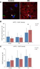
          Early life stress negatively impacts brain development and affects structure and function of parvalbumin immunopositive (PV+) inhibitory neurons. Main regulators of PV+ interneurons activity and plasticity are perineuronal nets (PNNs), an extracellular matrix formation that enwraps PV+ interneurons mainly in the neocortex and hippocampus. To experimentally address the impact of early life stress on the PNNs and PV+ interneurons in the medial prefrontal cortex and dorsal hippocampus in rats, we employed a 24 h maternal deprivation protocol. We show that maternal deprivation in the medial prefrontal cortex of adult rats caused a decrease in density of overall PNNs and PNNs that enwrap PV+ interneurons in the rostral cingulate cortex. Furthermore, a staining intensity decrease of overall PNNs and PNN+/PV+ cells was found in the prelimbic cortex. Finally, a decrease in both intensity and density of overall PNNs and PNNs surrounding PV+ cells was observed in the infralimbic cortex, together with increase in the intensity of VGAT inhibitory puncta. Surprisingly, maternal deprivation did not cause any changes in the density of PV+ interneurons in the mPFC, neither had it affected PNNs and PV+ interneurons in the hippocampus. Taken together, our findings indicate that PNNs, specifically the ones enwrapping PV+ interneurons in the medial prefrontal cortex, are affected by early life stress.

          Related collections

          Most cited references66

          • Record: found
          • Abstract: found
          • Article: not found

          Are the dorsal and ventral hippocampus functionally distinct structures?

          One literature treats the hippocampus as a purely cognitive structure involved in memory; another treats it as a regulator of emotion whose dysfunction leads to psychopathology. We review behavioral, anatomical, and gene expression studies that together support a functional segmentation into three hippocampal compartments: dorsal, intermediate, and ventral. The dorsal hippocampus, which corresponds to the posterior hippocampus in primates, performs primarily cognitive functions. The ventral (anterior in primates) relates to stress, emotion, and affect. Strikingly, gene expression in the dorsal hippocampus correlates with cortical regions involved in information processing, while genes expressed in the ventral hippocampus correlate with regions involved in emotion and stress (amygdala and hypothalamus).
            Bookmark
            • Record: found
            • Abstract: found
            • Article: not found

            Stress signalling pathways that impair prefrontal cortex structure and function.

            The prefrontal cortex (PFC) - the most evolved brain region - subserves our highest-order cognitive abilities. However, it is also the brain region that is most sensitive to the detrimental effects of stress exposure. Even quite mild acute uncontrollable stress can cause a rapid and dramatic loss of prefrontal cognitive abilities, and more prolonged stress exposure causes architectural changes in prefrontal dendrites. Recent research has begun to reveal the intracellular signalling pathways that mediate the effects of stress on the PFC. This research has provided clues as to why genetic or environmental insults that disinhibit stress signalling pathways can lead to symptoms of profound prefrontal cortical dysfunction in mental illness.
              Bookmark
              • Record: found
              • Abstract: found
              • Article: not found

              The effects of childhood maltreatment on brain structure, function and connectivity.

              Maltreatment-related childhood adversity is the leading preventable risk factor for mental illness and substance abuse. Although the association between maltreatment and psychopathology is compelling, there is a pressing need to understand how maltreatment increases the risk of psychiatric disorders. Emerging evidence suggests that maltreatment alters trajectories of brain development to affect sensory systems, network architecture and circuits involved in threat detection, emotional regulation and reward anticipation. This Review explores whether these alterations reflect toxic effects of early-life stress or potentially adaptive modifications, the relationship between psychopathology and brain changes, and the distinction between resilience, susceptibility and compensation.
                Bookmark

                Author and article information

                Contributors
                Journal
                Front Cell Dev Biol
                Front Cell Dev Biol
                Front. Cell Dev. Biol.
                Frontiers in Cell and Developmental Biology
                Frontiers Media S.A.
                2296-634X
                28 November 2022
                2022
                : 10
                : 982663
                Affiliations
                [1] 1 Center for Laser Microscopy , Institute for Physiology and Biochemistry “Jean Giaja” , Faculty of Biology , University of Belgrade , Belgrade, Serbia
                [2] 2 School of Medicine , Institute of Anatomy “Niko Miljanic” , University of Belgrade , Belgrade, Serbia
                [3] 3 Institut für Anatomie und Klinische Morphologie , Universität Witten/Herdecke , Witten, Germany
                [4] 4 Department of Neuroanatomy and Molecular Brain Research , Institute of Anatomy , Ruhr-Universität Bochum , Bochum, Germany
                [5] 5 Biology-Biochemistry Lab , School of Health Sciences , Faculty of Nursing , National and Kapodistrian University of Athens , Athens, Greece
                Author notes

                Edited by: Vladimir Sytnyk, University of New South Wales, Australia

                Reviewed by: Herbert Hildebrandt, Hannover Medical School, Germany

                Torkel Hafting, University of Oslo, Norway

                This article was submitted to Cell Adhesion and Migration, a section of the journal Frontiers in Cell and Developmental Biology

                Article
                982663
                10.3389/fcell.2022.982663
                9742529
                36518543
                98dad419-0887-42ab-a00a-ca2ddb65d935
                Copyright © 2022 Jakovljevic, Agatonovic, Aleksic, Aksic, Reiss, Förster, Stamatakis, Jakovcevski and Poleksic.

                This is an open-access article distributed under the terms of the Creative Commons Attribution License (CC BY). The use, distribution or reproduction in other forums is permitted, provided the original author(s) and the copyright owner(s) are credited and that the original publication in this journal is cited, in accordance with accepted academic practice. No use, distribution or reproduction is permitted which does not comply with these terms.

                History
                : 30 June 2022
                : 07 November 2022
                Categories
                Cell and Developmental Biology
                Original Research

                perineuronal nets,interneurons,maternal deprivation,early life stress,prefrontal cortex,hippocampus

                Comments

                Comment on this article