5
views
0
recommends
+1 Recommend
0 collections
    0
    shares
      • Record: found
      • Abstract: found
      • Article: found
      Is Open Access

      Reproducibility of a new classification of the anterior clinoid process of the sphenoid bone

      research-article

      Read this article at

      Bookmark
          There is no author summary for this article yet. Authors can add summaries to their articles on ScienceOpen to make them more accessible to a non-specialist audience.

          Abstract

          Background:

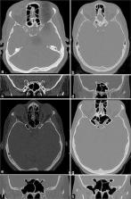
          Pneumatization of the anterior clinoid process (ACP) affects paraclinoid region surgery, this anatomical variation occurs in 6.6–27.7% of individuals, making its preoperative recognition essential given the need for correction based on the anatomy of the pneumatized process. This study was conducted to evaluate the reproducibility of an optic strut-based ACP pneumatization classification by presenting radiological examinations to a group of surgeons.

          Methods:

          Thirty cranial computer tomography (CT) scans performed from 2013 to 2014 were selected for analysis by neurosurgery residents and neurosurgeons. The evaluators received Google Forms with questionnaires on each scan, DICOM files to be manipulated in the Horos software for multiplanar reconstruction, and a collection of slides demonstrating the steps for classifying each type of ACP pneumatization. Interobserver agreement was calculated by the Fleiss kappa test.

          Results:

          Thirty CT scans were analyzed by 37 evaluators, of whom 20 were neurosurgery residents and 17 were neurosurgeons. The overall reproducibility of the ACP pneumatization classification showed a Fleiss kappa index of 0.49 (95% confidence interval: 0.49–0.50). The interobserver agreement indices for the residents and neurosurgeons were 0.52 (0.51–0.53) and 0.49 (0.48–0.50), respectively, and the difference was statistically significant ( P < 0.00001).

          Conclusion:

          The optic strut-based classification of ACP pneumatization showed acceptable concordance. Minor differences were observed in the agreement between the residents and neurosurgeons. These differences could be explained by the residents’ presumably higher familiarity with multiplanar reconstruction software.

          Related collections

          Most cited references23

          • Record: found
          • Abstract: not found
          • Book: not found

          Statistical methods for rates and proportions.

            Bookmark
            • Record: found
            • Abstract: found
            • Article: not found

            A combined epi- and subdural direct approach to carotid-ophthalmic artery aneurysms.

            A series of 14 patients with a carotid-ophthalmic artery aneurysm were treated operatively. In five patients the aneurysms were large, but only one of these had ruptured; four of these patients had symptoms of mass lesions. The remaining nine patients were operated on for a ruptured aneurysm; seven had subarachnoid hemorrhage due to the carotid-ophthalmic artery aneurysm and two had bleeding from another aneurysm in the presence of an asymptomatic carotid-ophthalmic artery aneurysm. All patients were treated by a combined epi- and subdural direct surgical approach, which excluded the carotid-ophthalmic artery aneurysm from the circulation and made possible the preservation of the adjacent structures. Two patients died: one a few hours after surgery from a massive thromboembolism of the pulmonary artery and another 2 months after surgery as a result of gastrointestinal bleeding. All the other patients showed postoperative improvement in symptoms and signs. This report focuses on a modified direct surgical approach involving exposure of the internal carotid artery proximal to the lesion, and of the ophthalmic artery, which is of primary importance in securing safe and complete occlusion of a carotid-ophthalmic artery aneurysm. Removal of individual bone structures at the base of the skull provides a better and safer exposure of the central segment of the internal carotid artery than does excessive and hazardous retraction of the brain.
              Bookmark
              • Record: found
              • Abstract: not found
              • Article: not found

              The cavernous sinus, the cavernous venous plexus, and the carotid collar.

                Bookmark

                Author and article information

                Contributors
                Journal
                Surg Neurol Int
                Surg Neurol Int
                Surgical Neurology International
                Scientific Scholar (USA )
                2229-5097
                2152-7806
                2020
                12 September 2020
                : 11
                : 281
                Affiliations
                [1 ]Department of Neurosurgery, Universidade Federal de São Paulo,
                [2 ]Department of Neurosurgery, Faculdade de Medicina de São Jose do Rio Preto, Sao Jose do Rio Preto,
                [3 ]Department of Neurosurgery, Centro Hospitalar Tondela-Viseu, EPE, Viseu, Portugal.
                [4 ]Department of Neurosurgery, Universidade de São Paulo, São Paulo, Brazil,
                Author notes
                [* ] Corresponding author: Marcos Devanir Silva da Costa, Department of Neurosurgery, Universidade Federal de São Paulo, São Paulo, Brazil. marcoscostaneuro@ 123456gmail.com
                Article
                SNI-11-281
                10.25259/SNI_133_2020
                7538961
                995a7338-cbeb-4712-8104-10a7e5ee1a49
                Copyright: © 2020 Surgical Neurology International

                This is an open-access article distributed under the terms of the Creative Commons Attribution-Non Commercial-Share Alike 4.0 License, which allows others to remix, tweak, and build upon the work non-commercially, as long as the author is credited and the new creations are licensed under the identical terms.

                History
                : 29 March 2020
                : 08 July 2020
                Categories
                Original Article

                Surgery
                anterior clinoid process,sphenoid bone,cavernous sinus,skull base,optic strut
                Surgery
                anterior clinoid process, sphenoid bone, cavernous sinus, skull base, optic strut

                Comments

                Comment on this article