13
views
0
recommends
+1 Recommend
0 collections
    0
    shares
      • Record: found
      • Abstract: found
      • Article: found
      Is Open Access

      Human Left Ventral Premotor Cortex Mediates Matching of Hand Posture to Object Use

      research-article

      Read this article at

      Bookmark
          There is no author summary for this article yet. Authors can add summaries to their articles on ScienceOpen to make them more accessible to a non-specialist audience.

          Abstract

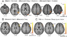
          Visuomotor transformations for grasping have been associated with a fronto-parietal network in the monkey brain. The human homologue of the parietal monkey region (AIP) has been identified as the anterior part of the intraparietal sulcus (aIPS), whereas the putative human equivalent of the monkey frontal region (F5) is located in the ventral part of the premotor cortex (vPMC). Results from animal studies suggest that monkey F5 is involved in the selection of appropriate hand postures relative to the constraints of the task. In humans, the functional roles of aIPS and vPMC appear to be more complex and the relative contribution of each region to grasp selection remains uncertain. The present study aimed to identify modulation in brain areas sensitive to the difficulty level of tool object - hand posture matching. Seventeen healthy right handed participants underwent fMRI while observing pictures of familiar tool objects followed by pictures of hand postures. The task was to decide whether the hand posture matched the functional use of the previously shown object. Conditions were manipulated for level of difficulty. Compared to a picture matching control task, the tool object – hand posture matching conditions conjointly showed increased modulation in several left hemispheric regions of the superior and inferior parietal lobules (including aIPS), the middle occipital gyrus, and the inferior temporal gyrus. Comparison of hard versus easy conditions selectively modulated the left inferior frontal gyrus with peak activity located in its opercular part (Brodmann area (BA) 44). We suggest that in the human brain, vPMC/BA44 is involved in the matching of hand posture configurations in accordance with visual and functional demands.

          Related collections

          Most cited references52

          • Record: found
          • Abstract: found
          • Article: not found

          Grasping objects: the cortical mechanisms of visuomotor transformation.

          Grasping requires coding of the object's intrinsic properties (size and shape), and the transformation of these properties into a pattern of distal (finger and wrist) movements. Computational models address this behavior through the interaction of perceptual and motor schemas. In monkeys, the transformation of an object's intrinsic properties into specific grips takes place in a circuit that is formed by the inferior parietal lobule and the inferior premotor area (area F5). Neurons in both these areas code size, shape and orientation of objects, and specific types of grip that are necessary to grasp them. Grasping movements are coded more globally in the inferior parietal lobule, whereas they are more segmented in area F5. In humans, neuropsychological studies of patients with lesions to the parietal lobule confirm that primitive shape characteristics of an object for grasping are analyzed in the parietal lobe, and also demonstrate that this 'pragmatic' analysis of objects is separated from the 'semantic' analysis performed in the temporal lobe.
            Bookmark
            • Record: found
            • Abstract: found
            • Article: not found

            Neuroimaging of cognitive functions in human parietal cortex.

            Functional neuroimaging has proven highly valuable in mapping human sensory regions, particularly visual areas in occipital cortex. Recent evidence suggests that human parietal cortex may also consist of numerous specialized subregions similar to those reported in neurophysiological studies of non-human primates. However, parietal activation generalizes across a wide variety of cognitive tasks and the extension of human brain mapping into higher-order "association cortex" may prove to be a challenge.
              Bookmark
              • Record: found
              • Abstract: found
              • Article: not found

              Goal representation in human anterior intraparietal sulcus.

              When a child reaches toward a cookie, the watching parent knows immediately what the child wants. The neural basis of this ability to interpret other people's actions in terms of their goals has been the subject of much speculation. Research with infants has shown that 6 month olds respond when they see an adult reach to a novel goal but habituate when an adult reaches to the same goal repeatedly. We used a similar approach in an event-related functional magnetic resonance imaging experiment. Adult participants observed a series of movies depicting goal-directed actions, with the sequence controlled so that some goals were novel and others repeated relative to the previous movie. Repeated presentation of the same goal caused a suppression of the blood oxygen level-dependent response in two regions of the left intraparietal sulcus. These regions were not sensitive to the trajectory taken by the actor's hand. This result demonstrates that the anterior intraparietal sulcus represents the goal of an observed action.
                Bookmark

                Author and article information

                Contributors
                Role: Editor
                Journal
                PLoS One
                PLoS ONE
                plos
                plosone
                PLoS ONE
                Public Library of Science (San Francisco, USA )
                1932-6203
                2013
                30 July 2013
                : 8
                : 7
                : e70480
                Affiliations
                [1 ]Department of Experimental Psychology, Ghent University, Ghent, Belgium
                [2 ]Ghent Institute for Functional and Metabolic Imaging, Ghent University, Ghent, Belgium
                [3 ]Department of Radiology, Ghent University, Ghent, Belgium
                Weill Cornell Medical College, United States of America
                Author notes

                Competing Interests: The authors have declared that no competing interests exist.

                Conceived and designed the experiments: GV JN PV. Performed the experiments: GV JN PH EV. Analyzed the data: GV JN. Contributed reagents/materials/analysis tools: PH EV PV. Wrote the paper: GV.

                Article
                PONE-D-13-11591
                10.1371/journal.pone.0070480
                3728237
                23936212
                9972883f-087c-404c-8084-860693911361
                Copyright @ 2013

                This is an open-access article distributed under the terms of the Creative Commons Attribution License, which permits unrestricted use, distribution, and reproduction in any medium, provided the original author and source are credited.

                History
                : 20 March 2013
                : 19 June 2013
                Page count
                Pages: 8
                Funding
                This study was supported by grants number G.0345.04 and G.0555.11 attributed to the first author by the Fund for Scientific Research – Flanders ( www.fwo.be). The funders had no role in study design, data collection and analysis, decision to publish, or preparation of the manuscript.
                Categories
                Research Article
                Biology
                Anatomy and Physiology
                Neurological System
                Motor Systems
                Neuroscience
                Cognitive Neuroscience
                Cognition
                Motor Reactions
                Neuroimaging
                Fmri
                Neurophysiology
                Motor Systems
                Behavioral Neuroscience
                Motor Systems
                Sensory Perception
                Medicine
                Anatomy and Physiology
                Neurological System
                Motor Systems
                Neurology
                Neuroimaging
                Social and Behavioral Sciences
                Psychology
                Sensory Perception

                Uncategorized
                Uncategorized

                Comments

                Comment on this article