0
views
0
recommends
+1 Recommend
0 collections
    0
    shares
      • Record: found
      • Abstract: found
      • Article: found
      Is Open Access

      Subcutaneous Fat Necrosis of Newborn: An Atypical Presentation

      letter
      ,
      Indian Journal of Dermatology
      Wolters Kluwer - Medknow

      Read this article at

      Bookmark
          There is no author summary for this article yet. Authors can add summaries to their articles on ScienceOpen to make them more accessible to a non-specialist audience.

          Abstract

          Sir, A full-term male baby weighing 3 kg born by vaginal delivery was referred at 18 h of age with respiratory distress. There was a history of perinatal asphyxia requiring resuscitation and meconium staining of liquor. In view of severe respiratory distress, the baby was intubated and ventilated. Chest X-ray showed few bilateral infiltrates suggestive of meconium aspiration syndrome. On day 4, the baby developed an erythematous rash over the back, and by the 7th day, it progressed to violaceous induration [Figures 1 and 2] Dermatology consultation was sought, and diagnosis of cellulitis was considered. On day 8, the infant had multiple brief episodes of convulsion managed with anticonvulsants. Cranial USG and MRI revealed intraventricular hemorrhage and bilateral periventricular changes consistent with perinatal hypoxia. By the 11th day, lesions progressed to multiple red, soft, fluctuant swellings, suggesting abscess formation. The lesions coalesced to form voluminous collection with inflamed overlying skin and surrounding area [Figure 3]. The baby was weaned off ventilator after 48 h. CBC at admission revealed Hb 14.9 g/dL, TLC 7200, platelets 61,000; CRP was negative. Blood counts on day 4 at manifestation of skin lesions showed drop in platelet count to 31,000 and CRP 6 mg/dl (0–8 mg/dl). Broad-spectrum antibiotics were commenced after sending blood culture as there was concern of sepsis. Ultrasonography showed hyperechoic subcutaneous tissue. Investigations on day 7 revealed Hb 15.2 g/dL, TLC 10,200, platelet 71,000, and quantitative CRP 158 mg/dL. Serum calcium was 10.6 mg/dl (8.8–11 mg/dl), ionic calcium 4.8 mmol/l (2–2.7 mmol/l), triglycerides 115 mg/dl. There was persistent thrombocytopenia on serial blood counts. USG at this point revealed increased echogenicity of subcutaneous fat with two loculated collections measuring 12 mm × 10 mm and 10 mm × 8 mm suggestive of subcutaneous fat necrosis with liquefaction. Blood cultures were sterile. Large collection necessitated needle aspiration, which was done under ultrasound guidance. Culture yielded no growth. There was white chalky pus-like discharge from the fluctuant 
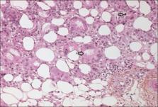
swellings for few days. Skin biopsy revealed subcutaneous tissue showing necrosis of adipocytes and infiltration by histiocytes and mononuclear cells. Some adipocytes showed needle-shaped clefts in radial arrangements, confirming the diagnosis of subcutaneous fat necrosis [Figure 4]. Platelet count and CRP normalized by 3 weeks. Calcium normalized by 4 weeks. Baby was discharged on day 34. The skin lesions regressed and complete resolution occurred around 6 weeks with scarring. Serum calcium repeated on the last follow-up at 3 months was normal. Figure 1 Erythematous lesion on the back Figure 2 Purple violaceous nodules and cystic swellings Figure 3 Large fluctuant collections Figure 4 Haematoxylin and eosin staining, ×400 showing A) Areas of necrosed adipocytes with needle shaped clefts B) Lipid laden histiocytes Subcutaneous fat necrosis is a form of benign lobular panniculitis affecting term or post-term neonates.[1 2 4] Leukocytosis, positive CRP, and low platelet count with cellulitis-like picture favored the initial diagnosis of neonatal sepsis in our case. Diagnosis of SCFN was first considered when ultrasonic evaluation revealed ill-defined areas of increased echogenicity in the subcutaneous fat with loculated collections and normal underlying muscles. Skin biopsy confirmed the diagnosis. USG with Doppler is a less invasive alternative to biopsy for diagnosis and shows high echo signal with or without calcifications and increased blood flow.[5] Possible differential diagnoses to be considered include bacterial cellulitis, erysipelas, sclerema neonatorum, and cold panniculitis.[3] In pyogenic abscess, the infant is often febrile or ill. SCFN can be a close mimic of cellulitis and sepsis; the clinical picture of an otherwise well child with a history of perinatal hypoxia is supportive. Diagnosis can be confirmed by skin biopsy showing characteristic findings of fat necrosis, radially arranged needle-shaped crystals in fat cells, granulomatous infiltrates composed of lymphocytes, macrophages, and giant cells. Clinicians should be aware of the association between SCFN and perinatal complications. Ultrasonography and Doppler studies can help diagnose this unusual entity and preclude unnecessary biopsy. Declaration of patient consent The authors certify that they have obtained all appropriate patient consent forms. In the form the patient(s) has/have given his/her/their consent for his/her/their images and other clinical information to be reported in the journal. The patients understand that their names and initials will not be published and due efforts will be made to conceal their identity, but anonymity cannot be guaranteed. Financial support and sponsorship Nil. Conflicts of interest There are no conflicts of interest.

          Related collections

          Most cited references5

          • Record: found
          • Abstract: found
          • Article: not found

          Subcutaneous fat necrosis of the newborn: a systematic evaluation of risk factors, clinical manifestations, complications and outcome of 16 children.

          Subcutaneous fat necrosis (SFN) of the newborn is a rare acute transient hypodermatitis that develops within the first weeks of life in term infants. It often follows a difficult delivery. Prognosis is generally good except for the development of hypercalcaemia in severe cases. Only several case reports or small patients series have been published. To evaluate risk factors, complications and outcomes of SFN in 16 consecutive patients seen from 1996 to 2002 in our Department of Paediatric Dermatology. On a case-report form created for the study, we recorded putative risk factors concerning the mother, pregnancy and delivery, clinical aspects of SFN, and early and late outcomes. The study was conducted in two stages: the first was a retrospective analysis of the observations and the second analysed data collected on children and their parents during a new consultation (n=10). All the children were born at term. Lesions appeared a mean of 4 days after delivery. Three-quarters of the children had diffuse SFN. Risk factors identified were newborn failure to thrive (12/16), forceps delivery (7/16), maternal high blood pressure (3/10) and/or diabetes (2/10), and newborn cardiac surgery (1/16). Putative novel risk factors were macrosomia (7/16), exposure to active (4/10) or passive (3/10) smoking during pregnancy, putative or known maternal, paternal or newborn risk factors for thrombosis (5/10), and dyslipidaemia (2/10). Complications were hypercalcaemia (9/16), pain (4/16), dyslipidaemia (1/16), renal insufficiency (1/16) and late subcutaneous atrophy (6/6). This study on 16 newborns with SFN provides new information. Familial or newborn risk factors for thrombosis are frequent. Macrosomia, familial dyslipidaemia and smoking should be evaluated. The main complications identified were severe pain, hypercalcaemia and subcutaneous atrophy.
            Bookmark
            • Record: found
            • Abstract: found
            • Article: not found

            Subcutaneous fat necrosis as a complication of whole-body cooling for birth asphyxia.

            Subcutaneous fat necrosis (SCFN) of the newborn is a form of panniculitis that affects full-term neonates who often have suffered either birth asphyxia or hypothermia. The induction of hypothermia in newborns is becoming frequently used to reduce the neurologic sequelae associated with birth asphyxia. The risk of SCFN in neonates undergoing this therapy is unknown. Observation We describe a neonate who developed an abscess-like presentation of SCFN and subsequent asymptomatic hypercalcemia after undergoing whole-body cooling for hypoxic-ischemic encephalopathy. Hypothermia protocols may be placing newborns at increased risk for the development of SCFN. Clinicians should recognize this association, and newborns who undergo therapeutic cooling should have frequent dermatologic assessments.
              Bookmark
              • Record: found
              • Abstract: found
              • Article: not found

              Subcutaneous fat necrosis of the newborn and associated hypercalcemia: A systematic review of the literature.

              Subcutaneous fat necrosis of the newborn is an uncommon disorder, and although usually benign, associated hypercalcemia can lead to complications such as failure to thrive and renal failure. Many sources suggest screening for hypercalcemia for 6 months following resolution of skin lesions, but little data are available to support this recommendation. This study examines existing published literature to better guide practitioners regarding screening evaluations of asymptomatic patients with subcutaneous fat necrosis. A systematic review of the literature was conducted using a PubMed English literature search. Data from case reports and case series were collected regarding the presence of hypercalcemia and associated complications, birth history, and age of onset/resolution of skin lesions and laboratory abnormalities. Approximately half (51%) of infants reported had hypercalcemia. Most (77%) developed detectable hypercalcemia within 30 days of skin lesion onset, and 95% developed detectable hypercalcemia within 60 days of skin lesion onset. Hypercalcemia was detected in only 4% of patients > 70 days following onset of skin lesions. Seventy-six percent had resolution of hypercalcemia within 4 weeks of detection. Hypercalcemia was more prevalent in full-term vs pre-term infants (P-value = 0.054), and higher birthweight was significantly associated with an increased risk of developing hypercalcemia (P-value = 0.022). Although gestational age trended toward significance, the only statistically significant clinical feature predicting the development of hypercalcemia was higher birthweight. Current recommendations for laboratory monitoring are not evidence-based, and this study provides interim data to guide practitioners until prospective, randomized controlled trials are conducted.
                Bookmark

                Author and article information

                Journal
                Indian J Dermatol
                Indian J Dermatol
                IJD
                Indian Journal of Dermatology
                Wolters Kluwer - Medknow (India )
                0019-5154
                1998-3611
                Mar-Apr 2022
                : 67
                : 2
                : 194-196
                Affiliations
                [1] From the Department of Paediatrics, Bharati Vidyapeeth (Deemed to be University) Medical College and Hospital, Sangli, Maharashtra, India E-mail: sarasubodh@ 123456yahoo.com
                Article
                IJD-67-194
                10.4103/ijd.ijd_550_21
                9455116
                9ab8f918-01c8-4c4c-8192-ad2c5d22df5b
                Copyright: © 2022 Indian Journal of Dermatology

                This is an open access journal, and articles are distributed under the terms of the Creative Commons Attribution-NonCommercial-ShareAlike 4.0 License, which allows others to remix, tweak, and build upon the work non-commercially, as long as appropriate credit is given and the new creations are licensed under the identical terms.

                History
                : June 2021
                : November 2021
                Categories
                Correspondence

                Dermatology
                Dermatology

                Comments

                Comment on this article