0
views
0
recommends
+1 Recommend
0 collections
    0
    shares
      • Record: found
      • Abstract: found
      • Article: found
      Is Open Access

      A Comparative Study of Immunofluorescence on Formalin-Fixed, Paraffin-Embedded Versus Fresh Frozen Kidney Biopsy

      research-article
      1 , 2 , 1 , , 1
      ,
      Cureus
      Cureus
      kidney disease, proteinase, antigen retrieval, formalin-fixed paraffin-embedded sections, direct immunofluorescence

      Read this article at

      Bookmark
          There is no author summary for this article yet. Authors can add summaries to their articles on ScienceOpen to make them more accessible to a non-specialist audience.

          Abstract

          Background

          Immunofluorescence techniques done on formalin-fixed, paraffin-embedded tissue can serve as salvage techniques in cases where immunofluorescence on the frozen section may not be adequate or available. The present study was undertaken to assess the diagnostic utility of paraffin immunofluorescence by proteinase K digestion on renal biopsy compared to fresh frozen immunofluorescence.

          Methodology

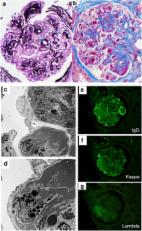
          The paraffin immunofluorescence by proteinase K digestion of paraffin-embedded renal biopsy (IF-FFPE) was standardized and compared with the immunofluorescence on fresh frozen tissue (IF-Frozen). A total of 50 cases of the native renal biopsy were included in the study, and their intensity for fluorescein isothiocyanate-labeled IgA, IgG, IgM, C3, kappa, and lambda was compared.

          Results

          A total of 50 cases of the native renal biopsy were included in the study, and their intensity for fluorescein isothiocyanate-labeled antibodies of IgA, IgG, IgM, C3, kappa, and lambda was compared. The difference of 2+ intensity of antibodies between IF-FFPE and IF-Frozen was noted mainly in lupus nephritis (15%), followed by IgA nephropathy (10%) and membranoproliferative glomerulonephritis (7%). IF-FFPE showed a sensitivity of 90.3%, 91.8%, 82.7%, 81.1%, 92.1%, and 94.6% for IgA, IgG, IgM, C3, kappa, and lambda, respectively, whereas specificity was 100% for IgA, IgG, C3, kappa, and lambda and 95.2% for IgM.

          Conclusions

          Immunofluorescence techniques done on formalin-fixed, paraffin-embedded tissue can serve as salvage techniques in kidney biopsies.

          Related collections

          Most cited references12

          • Record: found
          • Abstract: found
          • Article: not found

          Paraffin immunofluorescence in the renal pathology laboratory: more than a salvage technique.

          Immunofluorescence studies on paraffin-embedded tissue after Pronase digestion (paraffin immunofluorescence) is used as a salvage technique in renal pathology, when frozen tissue for routine immunofluorescence is inadequate. We have recently found that it is also useful in rare cases in which the immune deposits are 'masked' on routine immunofluorescence, giving false-negative staining by routine immunofluorescence and positive staining by paraffin immunofluorescence. This study aims to evaluate the role of paraffin immunofluorescence in clinical practice with emphasis on its utility to avoid misdiagnosis of cases with masked immune complex deposits. Paraffin immunofluorescence was used in 304 (6.1%) of 4969 native biopsies reviewed from our files. In 207 (68.1%) cases, paraffin immunofluorescence was used as a salvage technique. It was necessary for diagnosis in 24 (11.6%) and had a significant contribution in 63 (30.4%) of these cases. Paraffin immunofluorescence was used to evaluate masked deposits in 97 (31.9%) cases. In 61 (62.9%) of these cases it was used to evaluate masked immune complex glomerular deposits, and in 36 cases (37.1%) it was used to evaluate masked paraproteins. Of the cases where immune complex deposits were sought, paraffin immunofluorescence was necessary for diagnosis in 16 (26.2%) cases and had a significant contribution in 4 (6.6%) cases. Fourteen of the 20 cases with masked deposits had C3 dominant stain by routine immunofluorescence, which could have been misdiagnosed as C3 glomerulopathy. Overall, paraffin immunofluorescence was necessary or had a significant contribution to diagnosis in >1/3 of the cases and is a valuable technique in renal pathology.
            Bookmark
            • Record: found
            • Abstract: found
            • Article: not found

            Membranous-like glomerulopathy with masked IgG kappa deposits.

            The diagnostic classification of glomerulonephritis is determined by the interplay of changes seen using light, immunofluorescence, and electron microscopy of the renal biopsy. Routine direct immunofluorescence on fresh tissue is currently considered the gold standard for the detection and characterization of immune deposits. We recently found a peculiar form of glomerular immune complex deposition in which masked deposits required an antigen-retrieval step to be visualized. Over a 2-year period, 14 cases were characterized by numerous, large subepithelial deposits visualized by electron microscopy and C3-predominant staining by routine immunofluorescence on fresh tissue with weak to negative immunoglobulin staining. Repeat immunofluorescence after digestion of the formalin-fixed paraffin-embedded tissue with pronase elicited strong IgG-κ staining restricted within the deposits. The patients were often young with a mean age of 26 years and commonly had clinical evidence of vague autoimmune phenomenon. The clinicopathologic findings in this unusual form of glomerulopathy do not fit neatly into any currently existing diagnostic category. We have termed this unique form of glomerulopathy membranous-like glomerulopathy with masked IgG-κ deposits.
              Bookmark
              • Record: found
              • Abstract: found
              • Article: found
              Is Open Access

              Membranoproliferative glomerulonephritis with masked monotypic immunoglobulin deposits

              The diagnosis of membranoproliferative glomerulonephritis (MPGN) has recently undergone change from an electron microscopy-based classification scheme to one based largely on immunofluorescence findings. This change is due to the recognition that many of these cases are driven by abnormalities of the alternative complement cascade, resulting in the concept of C3 glomerulopathy. Here we reviewed our case files to identify those with an MPGN pattern that show false negative staining for monoclonal immunoglobulins by routine immunofluorescence. Monoclonal immunoglobulin deposits were unmasked by performing immunofluorescence on formalin-fixed paraffin embedded tissue after protease digestion. Clinico-pathological details of 16 such cases with a mean serum creatinine of 2.7 mg/dl and mean 24 h proteinuria of 7.1 g were then determined. Hypocomplementemia was present in two-thirds of patients. Fourteen patients had a paraprotein on serum immunofixation, all of which matched the biopsy immunofluorescence staining pattern. Bone marrow biopsy showed plasma cell dyscrasia or B-cell lymphoproliferative disorder in 13 patients. Ten of these patients had findings on biopsy most consistent with C3 glomerulonephritis prior to performing paraffin immunofluorescence. Thus a high index of suspicion is necessary to avoid misdiagnosis in these cases, as many would have been mistakenly diagnosed as C3 glomerulopathy or unclassified MPGN if paraffin immunofluorescence was not performed.
                Bookmark

                Author and article information

                Journal
                Cureus
                Cureus
                2168-8184
                Cureus
                Cureus (Palo Alto (CA) )
                2168-8184
                26 June 2023
                June 2023
                : 15
                : 6
                : e40978
                Affiliations
                [1 ] Pathology, North Eastern Indira Gandhi Regional Institute of Health and Medical Sciences (NEIGRIHMS), Shillong, IND
                [2 ] Allied Health Sciences, Martin Luther Christian University, Shillong, IND
                Author notes
                Article
                10.7759/cureus.40978
                10370476
                9bd2bae5-bb74-4167-8cd5-f92cd37f84e1
                Copyright © 2023, Das et al.

                This is an open access article distributed under the terms of the Creative Commons Attribution License, which permits unrestricted use, distribution, and reproduction in any medium, provided the original author and source are credited.

                History
                : 26 June 2023
                Categories
                Pathology
                Allergy/Immunology
                Nephrology

                kidney disease,proteinase,antigen retrieval,formalin-fixed paraffin-embedded sections,direct immunofluorescence

                Comments

                Comment on this article