21
views
0
recommends
+1 Recommend
0 collections
    0
    shares
      • Record: found
      • Abstract: found
      • Article: found
      Is Open Access

      Quantitative three-dimensional myocardial perfusion cardiovascular magnetic resonance with accurate two-dimensional arterial input function assessment

      research-article

      Read this article at

      Bookmark
          There is no author summary for this article yet. Authors can add summaries to their articles on ScienceOpen to make them more accessible to a non-specialist audience.

          Abstract

          Background

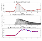
          Quantification of myocardial perfusion from first-pass cardiovascular magnetic resonance (CMR) images at high contrast agent (CA) dose requires separate acquisition of blood pool and myocardial tissue enhancement. In this study, a dual-sequence approach interleaving 2D imaging of the arterial input function with high-resolution 3D imaging for myocardial perfusion assessment is presented and validated for low and high CA dose.

          Methods

          A dual-sequence approach interleaving 2D imaging of the aortic root and 3D imaging of the whole left ventricle using highly accelerated k-t PCA was implemented. Rest perfusion imaging was performed in ten healthy volunteers after administration of a Gadolinium-based CA at low (0.025 mmol/kg b.w.) and high dose (0.1 mmol/kg b.w.). Arterial input functions extracted from the 2D and 3D images were analysed for both doses. Myocardial contrast-to-noise ratios (CNR) were compared across volunteers and doses. Variations of myocardial perfusion estimates between volunteers and across myocardial territories were studied.

          Results

          High CA dose imaging resulted in strong non-linearity of the arterial input function in the 3D images at peak CA concentration, which was avoided when the input function was derived from the 2D images. Myocardial CNR was significantly increased at high dose compared to low dose, with a 2.6-fold mean CNR gain. Most robust myocardial blood flow estimation was achieved using the arterial input function extracted from the 2D image at high CA dose. In this case, myocardial blood flow estimates varied by 24 % between volunteers and by 20 % between myocardial territories when analysed on a per-volunteer basis.

          Conclusion

          Interleaving 2D imaging for arterial input function assessment enables robust quantitative 3D myocardial perfusion imaging at high CA dose.

          Related collections

          Most cited references44

          • Record: found
          • Abstract: found
          • Article: not found

          k-t PCA: temporally constrained k-t BLAST reconstruction using principal component analysis.

          The k-t broad-use linear acquisition speed-up technique (BLAST) has become widespread for reducing image acquisition time in dynamic MRI. In its basic form k-t BLAST speeds up the data acquisition by undersampling k-space over time (referred to as k-t space). The resulting aliasing is resolved in the Fourier reciprocal x-f space (x = spatial position, f = temporal frequency) using an adaptive filter derived from a low-resolution estimate of the signal covariance. However, this filtering process tends to increase the reconstruction error or lower the achievable acceleration factor. This is problematic in applications exhibiting a broad range of temporal frequencies such as free-breathing myocardial perfusion imaging. We show that temporal basis functions calculated by subjecting the training data to principal component analysis (PCA) can be used to constrain the reconstruction such that the temporal resolution is improved. The presented method is called k-t PCA.
            Bookmark
            • Record: found
            • Abstract: found
            • Article: found
            Is Open Access

            Quantification of myocardial perfusion by cardiovascular magnetic resonance

            The potential of contrast-enhanced cardiovascular magnetic resonance (CMR) for a quantitative assessment of myocardial perfusion has been explored for more than a decade now, with encouraging results from comparisons with accepted "gold standards", such as microspheres used in the physiology laboratory. This has generated an increasing interest in the requirements and methodological approaches for the non-invasive quantification of myocardial blood flow by CMR. This review provides a synopsis of the current status of the field, and introduces the reader to the technical aspects of perfusion quantification by CMR. The field has reached a stage, where quantification of myocardial perfusion is no longer a claim exclusive to nuclear imaging techniques. CMR may in fact offer important advantages like the absence of ionizing radiation, high spatial resolution, and an unmatched versatility to combine the interrogation of the perfusion status with a comprehensive tissue characterization. Further progress will depend on successful dissemination of the techniques for perfusion quantification among the CMR community.
              Bookmark
              • Record: found
              • Abstract: found
              • Article: not found

              Magnetic resonance quantification of the myocardial perfusion reserve with a Fermi function model for constrained deconvolution.

              The myocardial perfusion reserve, defined as the ratio of hyperemic and basal myocardial blood flow, is a useful indicator of the functional significance of a coronary artery lesion. Rapid magnetic resonance (MR) imaging for the noninvasive detection of a bolus-injected contrast agent as a MR tracer is applied to the measurement of regional tissue perfusion during rest and hyperemia, in patients with microvascular dysfunction. A Fermi function model for the distribution of tracer residence times in the myocardium is used to fit the MR signal curves. The myocardial perfusion reserve is calculated from the impulse response amplitudes for rest and hyperemia. The assumptions of the model are tested with Monte Carlo simulations, using a multiple path, axially distributed mathematical model of blood tissue exchange, which allows for systematic variation of blood flow, vascular volume, and capillary permeability. For a contrast-to-noise ratio of 6:1, and over a range of flows from 0.5 to 4.0 ml/min per g of tissue, the ratio of the impulse response amplitudes for hyperemic and basal flows is linearly proportional to the ratio of model blood flows, if the mean transit time of the input function is shorter than approximately 9 s. The uncertainty in the blood flow reserve estimates grows both at low ( 3-4 ml/min/g) flows. The predictions of the Monte Carlo simulations agree with the results of MR first pass studies in patients without significant coronary artery lesions and microvascular dysfunction, where the perfusion reserve in the territory of the left anterior descending coronary artery (LAD) correlates linearly with the intracoronary Doppler ultrasound flow reserve in the LAD (r = 0.84), in agreement with previous PET studies.
                Bookmark

                Author and article information

                Contributors
                wissmann@biomed.ee.ethz.ch
                markus.niemann@hs-furtwangen.de
                alexander.gotschy@usz.ch
                robert.manka@usz.ch
                + 41 44 632 3549 , kozerke@biomed.ee.ethz.ch
                Journal
                J Cardiovasc Magn Reson
                J Cardiovasc Magn Reson
                Journal of Cardiovascular Magnetic Resonance
                BioMed Central (London )
                1097-6647
                1532-429X
                4 December 2015
                4 December 2015
                2015
                : 17
                : 108
                Affiliations
                [ ]Institute for Biomedical Engineering, University and ETH Zurich, Gloriastrasse 35, 8092 Zurich, Switzerland
                [ ]Clinic of Cardiology, University Hospital Zurich, Zurich, Switzerland
                [ ]Furtwangen University, Faculty Mechanical and Medical Engineering, Villingen-Schwenningen, Germany
                [ ]Department of Internal Medicine, University Hospital Zurich, Zurich, Switzerland
                [ ]Institute of Diagnostic and Interventional Radiology, University Hospital Zurich, Zurich, Switzerland
                [ ]Imaging Sciences and Biomedical Engineering, King’s College London, London, UK
                Article
                212
                10.1186/s12968-015-0212-3
                4669617
                26637221
                9c83d490-8d82-4ddf-9a28-9c6ff3a593eb
                © Wissmann et al. 2015

                Open AccessThis article is distributed under the terms of the Creative Commons Attribution 4.0 International License ( http://creativecommons.org/licenses/by/4.0/), which permits unrestricted use, distribution, and reproduction in any medium, provided you give appropriate credit to the original author(s) and the source, provide a link to the Creative Commons license, and indicate if changes were made. The Creative Commons Public Domain Dedication waiver ( http://creativecommons.org/publicdomain/zero/1.0/) applies to the data made available in this article, unless otherwise stated.

                History
                : 20 April 2015
                : 24 November 2015
                Categories
                Research
                Custom metadata
                © The Author(s) 2015

                Cardiovascular Medicine
                dce-mri,first-pass myocardial perfusion imaging,arterial input function,myocardial blood flow estimates,dual-sequence imaging

                Comments

                Comment on this article