45
views
0
recommends
+1 Recommend
0 collections
    0
    shares
      • Record: found
      • Abstract: found
      • Article: found
      Is Open Access

      Trpm4 Gene Invalidation Leads to Cardiac Hypertrophy and Electrophysiological Alterations

      research-article

      Read this article at

      Bookmark
          There is no author summary for this article yet. Authors can add summaries to their articles on ScienceOpen to make them more accessible to a non-specialist audience.

          Abstract

          Rationale

          TRPM4 is a non-selective Ca 2+-activated cation channel expressed in the heart, particularly in the atria or conduction tissue. Mutations in the Trpm4 gene were recently associated with several human conduction disorders such as Brugada syndrome. TRPM4 channel has also been implicated at the ventricular level, in inotropism or in arrhythmia genesis due to stresses such as ß-adrenergic stimulation, ischemia-reperfusion, and hypoxia re-oxygenation. However, the physiological role of the TRPM4 channel in the healthy heart remains unclear.

          Objectives

          We aimed to investigate the role of the TRPM4 channel on whole cardiac function with a Trpm4 gene knock-out mouse ( Trpm4 -/-) model.

          Methods and Results

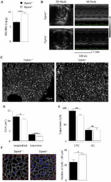
          Morpho-functional analysis revealed left ventricular (LV) eccentric hypertrophy in Trpm4 -/- mice, with an increase in both wall thickness and chamber size in the adult mouse (aged 32 weeks) when compared to Trpm4 +/+ littermate controls. Immunofluorescence on frozen heart cryosections and qPCR analysis showed no fibrosis or cellular hypertrophy. Instead, cardiomyocytes in Trpm4 -/- mice were smaller than Trpm4 +/+ with a higher density. Immunofluorescent labeling for phospho-histone H3, a mitosis marker, showed that the number of mitotic myocytes was increased 3-fold in the Trpm4 -/- neonatal stage, suggesting hyperplasia. Adult Trpm4 -/- mice presented multilevel conduction blocks, as attested by PR and QRS lengthening in surface ECGs and confirmed by intracardiac exploration. Trpm4 -/- mice also exhibited Luciani-Wenckebach atrioventricular blocks, which were reduced following atropine infusion, suggesting paroxysmal parasympathetic overdrive. In addition, Trpm4 -/- mice exhibited shorter action potentials in atrial cells. This shortening was unrelated to modifications of the voltage-gated Ca 2+ or K + currents involved in the repolarizing phase.

          Conclusions

          TRPM4 has pleiotropic roles in the heart, including the regulation of conduction and cellular electrical activity which impact heart development.

          Related collections

          Most cited references48

          • Record: found
          • Abstract: found
          • Article: not found

          Rapid transition of cardiac myocytes from hyperplasia to hypertrophy during postnatal development.

          The switch from myocyte hyperplasia to hypertrophy occurs during the early postnatal period. The exact temporal sequence when cardiac myocytes cease dividing and become terminally differentiated is not certain, although it is currently believed that the transition takes place gradually over a 1-2-week period. The present investigation has characterized the growth pattern of cardiac myocytes during the early postnatal period. Cardiac myocytes were enzymatically isolated from the hearts of 1, 2, 3, 4, 6, 8, 10, and 12-day-old rats for the measurements of binucleation, cell volume and myocyte number. Almost all myocytes were mononucleated and cell volume remained relatively constant during the first 3 days of age. Increases in cell volume and binucleation of myocytes were first detected at day 4. Myocyte volume increased 2.5-fold from day 3 to day 12 (1416 +/- 320 compared to 3533 +/- 339 microns 3). The percentage of binucleated myocytes began to increase at day 4 and proceeded at a high rate, reaching the adult level of approximately 90% at day 12. Myocyte number increased 68% during the first 3 days (from 13.6 +/- 3.5 x 10(6) at day 1 to 22.9 +/- 5.6 x 10(10) at day 3) and remained constant thereafter. To confirm that no further myocyte division exists after 4 days, bromodeoxyuridine (Brdu) was administered to 4-day-old rats and the fate of DNA-synthesizing myocytes was examined 2 h and 2, 4, 6 and 8 days after Brdu injection. About 12% of myocytes were labeled with Brdu at 2 h and all were mononucleated at that time. Gradually, these Brdu-labeled myocytes became binucleated. However, the percentage of labeled myocytes in all groups was identical, indicating that DNA-synthesizing myocytes were becoming binucleated without further cell division after 4 days of age. Within 8 days after injection, approximately 82% of total labeled myocytes were binucleated, while the others remained mononucleated. Sarcomeric alpha-actinin was fully disassembled in dividing myocytes of 2-day-old rats, while typical alpha-actinin striations were present in dividing myocytes of 4-day-old rats. The results from this study suggest that a rapid switch from myocyte hyperplasia to hypertrophy occurs between postnatal day 3 and 4 in rat hearts.
            Bookmark
            • Record: found
            • Abstract: found
            • Article: not found

            TRP-PLIK, a bifunctional protein with kinase and ion channel activities.

            We cloned and characterized a protein kinase and ion channel, TRP-PLIK. As part of the long transient receptor potential channel subfamily implicated in control of cell division, it is a protein that is both an ion channel and a protein kinase. TRP-PLIK phosphorylated itself, displayed a wide tissue distribution, and, when expressed in CHO-K1 cells, constituted a nonselective, calcium-permeant, 105-picosiemen, steeply outwardly rectifying conductance. The zinc finger containing alpha-kinase domain was functional. Inactivation of the kinase activity by site-directed mutagenesis and the channel's dependence on intracellular adenosine triphosphate (ATP) demonstrated that the channel's kinase activity is essential for channel function.
              Bookmark
              • Record: found
              • Abstract: found
              • Article: not found

              Cardiomyocyte DNA synthesis and binucleation during murine development.

              Cardiomyocyte DNA synthesis and binucleation indexes were determined during murine development. Cardiomyocyte DNA synthesis occurred in two temporally distinct phases. The first phase occurred during fetal life and was associated exclusively with cardiomyocyte proliferation. The second phase occurred during early neonatal life and was associated with binucleation. Collectively, these results suggest that cardiomyocyte reduplication ceases during late fetal life. Northern and Western blot analyses identified several candidate genes that were differentially expressed during the reduplicative and binucleation phases of cardiomyocyte growth.
                Bookmark

                Author and article information

                Contributors
                Role: Editor
                Journal
                PLoS One
                PLoS ONE
                plos
                plosone
                PLoS ONE
                Public Library of Science (San Francisco, USA )
                1932-6203
                2014
                22 December 2014
                : 9
                : 12
                : e115256
                Affiliations
                [1 ]INSERM U1046, Université Montpellier1, Université Montpellier2, Montpellier, France
                [2 ]Equipe Avenir, INSERM U1149, Université Paris 7, Paris, France
                [3 ]CHRU Montpellier, Service de Cardiologie, Montpellier, France
                Georgia State University, United States of America
                Author notes

                Competing Interests: The authors have declared that no competing interests exist.

                Conceived and designed the experiments: MD JT SR. Performed the experiments: MD JT M. Gueffier ZK CC NS FA. Analyzed the data: MD JT M. Gueffier ZK CC NS FA. Contributed reagents/materials/analysis tools: MD JT PL SR. Wrote the paper: MD JT AF FA M. Granier JLP PL SR.

                Article
                PONE-D-14-31075
                10.1371/journal.pone.0115256
                4274076
                25531103
                a349e69b-7795-40f7-b0c7-3fe3d2fd1396
                Copyright @ 2014

                This is an open-access article distributed under the terms of the Creative Commons Attribution License, which permits unrestricted use, distribution, and reproduction in any medium, provided the original author and source are credited.

                History
                : 17 July 2014
                : 20 November 2014
                Page count
                Pages: 28
                Funding
                MD SR, grant from Université Montpellier 2; PL MD SR, grant from Agence Nationale de la Recherche (“Target channel”; RPV09046FSA); SR, grant from Agence Nationale de la Recherche ("Beat-genesis" 2010 blanc 1128 03"); SR, grant from Région Languedoc-Roussillon (GEPETOS). The funders had no role in study design, data collection and analysis, decision to publish, or preparation of the manuscript.
                Categories
                Research Article
                Biology and Life Sciences
                Anatomy
                Cardiovascular Anatomy
                Heart
                Ventricles (Heart)
                Biochemistry
                Proteins
                Ion Channels
                Transient Receptor Potential Channels
                Neuroscience
                Neurophysiology
                Action Potentials
                Physiology
                Electrophysiology
                Membrane Potential
                Cardiovascular Physiology
                Medicine and Health Sciences
                Cardiology
                Cardiac Hypertrophy
                Research and Analysis Methods
                Model Organisms
                Animal Models
                Mouse Models
                Custom metadata
                The authors confirm that all data underlying the findings are fully available without restriction. All relevant data are within the paper and its Supporting Information files.

                Uncategorized
                Uncategorized

                Comments

                Comment on this article