7
views
0
recommends
+1 Recommend
0 collections
    0
    shares
      • Record: found
      • Abstract: found
      • Article: found
      Is Open Access

      Rare Coronary Embolism Secondary to Cardioversion of Atrial Fibrillation

      case-report

      Read this article at

      Bookmark
          There is no author summary for this article yet. Authors can add summaries to their articles on ScienceOpen to make them more accessible to a non-specialist audience.

          Abstract

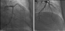
          The diagnosis and management of myocardial infarction with nonobstructive coronary arteries (MINOCA) are difficult due to its variable presentations, different causes, and challenging diagnostic approaches. Cardiac imaging modalities including cardiac magnetic resonance (CMR) are very useful tools for diagnosing and managing MINOCA. Myocardial infarction (MI) can be caused by coronary emboli that can be contributed to atrial fibrillation (AF). Rarely, coronary embolism with resultant MINOCA can occur after direct current cardioversion (DCCV) even in fully anticoagulated patients. We present a rare case of a coronary embolism following DCCV as well as a CMR finding of microvascular obstruction (MVO), which has not previously been reported after DCCV. This case also emphasizes the value of obtaining a CMR for patients with MINOCA.

          Related collections

          Most cited references10

          • Record: found
          • Abstract: not found
          • Article: not found

          ESC working group position paper on myocardial infarction with non-obstructive coronary arteries

            Bookmark
            • Record: found
            • Abstract: found
            • Article: not found

            Microvascular obstruction: underlying pathophysiology and clinical diagnosis.

            Successful restoration of epicardial coronary artery patency after prolonged occlusion might result in microvascular obstruction (MVO) and is observed both experimentally as well as clinically. In reperfused myocardium, myocytes appear edematous and swollen from osmotic overload. Endothelial cell changes usually accompany the alterations seen in myocytes but lag behind myocardial cell injury. Endothelial cells become voluminous, with large intraluminal endothelial protrusions into the vascular lumen, and together with swollen surrounding myocytes occlude capillaries. The infiltration and activation of neutrophils and platelets and the deposition of fibrin also play an important role in reperfusion-induced microvascular damage and obstruction. In addition to these ischemia-reperfusion-related events, coronary microembolization of atherosclerotic debris after percutaneous coronary intervention is responsible for a substantial part of clinically observed MVO. Microvascular flow after reperfusion is spatially and temporally complex. Regions of hyperemia, impaired vasodilatory flow reserve and very low flow coexist and these perfusion patterns vary over time as a result of reperfusion injury. The MVO first appears centrally in the infarct core extending toward the epicardium over time. Accurate detection of MVO is crucial, because it is independently associated with adverse ventricular remodeling and patient prognosis. Several techniques (coronary angiography, myocardial contrast echocardiography, cardiovascular magnetic resonance imaging, electrocardiography) measuring slightly different biological and functional parameters are used clinically and experimentally. Currently there is no consensus as to how and when MVO should be evaluated after acute myocardial infarction. Copyright (c) 2010 American College of Cardiology Foundation. Published by Elsevier Inc. All rights reserved.
              Bookmark
              • Record: found
              • Abstract: found
              • Article: not found

              MR imaging of myocardial infarction.

              Magnetic resonance (MR) imaging plays an important role in evaluation of various aspects of myocardial infarction (MI). MR imaging is useful in establishing the diagnosis of acute MI, particularly in patients who present with symptoms of MI but outside the diagnostic time frame of altered cardiac enzyme levels or with clinical features of acute MI but without an angiographic culprit lesion. MR imaging is valuable in establishing a diagnosis of chronic MI and distinguishing this condition from nonischemic cardiomyopathies, mainly through use of delayed-enhancement patterns. MR imaging also provides clinicians with several prognostic indicators that enable risk stratification, such as scar burden, microvascular obstruction, hemorrhage, and peri-infarct ischemia. The extent and transmurality of scar burden have been shown to have independent and incremental prognostic power over a range of left ventricular function. The extent of scarring at MR imaging is an important predictor of successful outcome after revascularization procedures, and extensive scarring in the lateral wall indicates poor outcome after cardiac resynchronization therapy. Scar size at MR imaging is also a useful surrogate end point in clinical trials. Finally, MR imaging can be used to detect complications of MI, such as aneurysms, pericarditis, ventricular septal defect, thrombus, and mitral regurgitation. Supplemental material available at http://radiographics.rsna.org/lookup/suppl/doi:10.1148/rg.335125722/-/DC1.
                Bookmark

                Author and article information

                Journal
                Cureus
                Cureus
                2168-8184
                Cureus
                Cureus (Palo Alto (CA) )
                2168-8184
                21 April 2022
                April 2022
                : 14
                : 4
                : e24354
                Affiliations
                [1 ] Cardiology, The University of Tennessee Health Science Center, Memphis, USA
                [2 ] Cardiology, Methodist University Hospital, Methodist Le Bonheur Healthcare/The University of Tennessee Health Science Center, Memphis, USA
                Author notes
                Article
                10.7759/cureus.24354
                9124055
                35611046
                a4c55c5d-acdf-423e-ba8b-9e1832b09dfb
                Copyright © 2022, Alkhatib et al.

                This is an open access article distributed under the terms of the Creative Commons Attribution License, which permits unrestricted use, distribution, and reproduction in any medium, provided the original author and source are credited.

                History
                : 21 April 2022
                Categories
                Cardiology
                Internal Medicine
                Radiology

                minoca,coronary embolism,microvascular obstruction (mvo),direct current cardioversion (dccv),atrial fibrillation (af),coronary artery embolism,cardiac magnetic resonance (cmr),myocardial infarction with non-obstructive coronary arteries (minoca)

                Comments

                Comment on this article