7
views
0
recommends
+1 Recommend
0 collections
    0
    shares
      • Record: found
      • Abstract: found
      • Article: found
      Is Open Access

      K Ca3.1 Channels Confer Radioresistance to Breast Cancer Cells

      research-article

      Read this article at

      Bookmark
          There is no author summary for this article yet. Authors can add summaries to their articles on ScienceOpen to make them more accessible to a non-specialist audience.

          Abstract

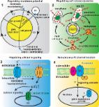
          K Ca3.1 K + channels reportedly contribute to the proliferation of breast tumor cells and may serve pro-tumor functions in the microenvironment. The putative interaction of K Ca3.1 with major anti-cancer treatment strategies, which are based on cytotoxic drugs or radiotherapy, remains largely unexplored. We employed K Ca3.1-proficient and -deficient breast cancer cells derived from breast cancer-prone MMTV-PyMT mice, pharmacological K Ca3.1 inhibition, and a syngeneic orthotopic mouse model to study the relevance of functional K Ca3.1 for therapy response. The K Ca3.1 status of MMTV-PyMT cells did not determine tumor cell proliferation after treatment with different concentrations of docetaxel, doxorubicin, 5-fluorouracil, or cyclophosphamide. K Ca3.1 activation by ionizing radiation (IR) in breast tumor cells in vitro, however, enhanced radioresistance, probably via an involvement of the channel in IR-stimulated Ca 2+ signals and DNA repair pathways. Consistently, K Ca3.1 knockout increased survival time of wildtype mice upon syngeneic orthotopic transplantation of MMTV-PyMT tumors followed by fractionated radiotherapy. Combined, our results imply that K Ca3.1 confers resistance to radio- but not to chemotherapy in the MMTV-PyMT breast cancer model. Since K Ca3.1 is druggable, K Ca3.1 targeting concomitant to radiotherapy seems to be a promising strategy to radiosensitize breast tumors.

          Related collections

          Most cited references43

          • Record: found
          • Abstract: found
          • Article: not found

          Ca2+ signalling checkpoints in cancer: remodelling Ca2+ for cancer cell proliferation and survival.

          Increases in cytosolic free Ca2+ ([Ca2+]i) represent a ubiquitous signalling mechanism that controls a variety of cellular processes, including proliferation, metabolism and gene transcription, yet under certain conditions increases in intracellular Ca2+ are cytotoxic. Thus, in using Ca2+ as a messenger, cells walk a tightrope in which [Ca2+]i is strictly maintained within defined boundaries. To adhere to these boundaries and to sustain their modified phenotype, many cancer cells remodel the expression or activity of their Ca2+ signalling apparatus. Here, we review the role of Ca2+ in promoting cell proliferation and cell death, how these processes are remodelled in cancer and the opportunities this might provide for therapeutic intervention.
            Bookmark
            • Record: found
            • Abstract: found
            • Article: found
            Is Open Access

            Targeting potassium channels in cancer

            Potassium channels are pore-forming transmembrane proteins that regulate a multitude of biological processes by controlling potassium flow across cell membranes. Aberrant potassium channel functions contribute to diseases such as epilepsy, cardiac arrhythmia, and neuromuscular symptoms collectively known as channelopathies. Increasing evidence suggests that cancer constitutes another category of channelopathies associated with dysregulated channel expression. Indeed, potassium channel–modulating agents have demonstrated antitumor efficacy. Potassium channels regulate cancer cell behaviors such as proliferation and migration through both canonical ion permeation–dependent and noncanonical ion permeation–independent functions. Given their cell surface localization and well-known pharmacology, pharmacological strategies to target potassium channel could prove to be promising cancer therapeutics.
              Bookmark
              • Record: found
              • Abstract: found
              • Article: not found

              Improvements in haemolysis and indicators of erythrocyte survival do not correlate with acute vaso-occlusive crises in patients with sickle cell disease: a phase III randomized, placebo-controlled, double-blind study of the Gardos channel blocker senicapoc (ICA-17043).

              Red blood cell (RBC) hydration is regulated in part by the Ca(2+) -activated K(+) efflux (Gardos) channel. Senicapoc selectively blocks potassium efflux through the Gardos channel, reducing RBC dehydration and haemolysis, and increasing haemoglobin levels in sickle cell disease (SCD). This randomized, placebo-controlled trial was designed to determine the safety and clinical efficacy of senicapoc in SCD patients. One hundred and forty-five patients were randomized to receive senicapoc and 144 patients to receive placebo for 52 weeks. Consistent with a previous study, patients in the senicapoc group had significantly increased haematocrit, haemoglobin, and decreased numbers of both dense erythrocytes and reticulocytes when compared to the placebo group. The unblinded Data Monitoring Committee terminated this study early due to a lack of efficacy when it determined that, despite improvements in anaemia and haemolysis, no significant improvement in the rate of sickle cell painful crises was observed in patients treated with senicapoc compared to those on placebo (0·38 vs. 0·31, respectively). Comparisons of the times to first, second and third crises between the senicapoc and placebo groups were not statistically significant. Nausea and urinary tract infections occurred more frequently in the senicapoc group than placebo. Serious adverse events were similar in the two groups. © 2011 Blackwell Publishing Ltd.
                Bookmark

                Author and article information

                Journal
                Cancers (Basel)
                Cancers (Basel)
                cancers
                Cancers
                MDPI
                2072-6694
                01 September 2019
                September 2019
                : 11
                : 9
                : 1285
                Affiliations
                [1 ]Department of Pharmacology, Toxicology and Clinical Pharmacy, Institute of Pharmacy, University of Tuebingen, 72076 Tuebingen, Germany
                [2 ]Dr Margarete Fischer-Bosch-Institute of Clinical Pharmacology, 70376 Stuttgart and University of Tuebingen, 72076 Tuebingen, Germany
                [3 ]Department of Radiation Oncology, University of Tuebingen, 72076 Tuebingen, Germany
                Author notes
                [* ]Correspondence: stephan.huber@ 123456uni-tuebingen.de (S.M.H.); robert.lukowski@ 123456uni-tuebingen.de (R.L.); Tel.: +49-7071-29-82183 (S.M.H.); +49-7071-29-74550 (R.L.); Fax: +49-7071-29-4944 (S.M.H.); +49-7071-29-2476 (R.L.)
                Author information
                https://orcid.org/0000-0002-6741-7658
                Article
                cancers-11-01285
                10.3390/cancers11091285
                6770875
                31480522
                a75ee3e6-69bf-4b2c-afa6-bfae2b3d20cc
                © 2019 by the authors.

                Licensee MDPI, Basel, Switzerland. This article is an open access article distributed under the terms and conditions of the Creative Commons Attribution (CC BY) license ( http://creativecommons.org/licenses/by/4.0/).

                History
                : 02 May 2019
                : 21 August 2019
                Categories
                Article

                breast cancer,ionizing radiation,intermediate conductance calcium-activated potassium channel,kca3.1,sk4,ik,kcnn4,mouse mammary tumor virus polyoma middle t antigen,mmtv-pymt,tram-34

                Comments

                Comment on this article