3
views
0
recommends
+1 Recommend
0 collections
    0
    shares
      • Record: found
      • Abstract: found
      • Article: found
      Is Open Access

      Surgical treatment of a rare rosette-forming glioneuronal tumor in the pineal region

      case-report

      Read this article at

      Bookmark
          There is no author summary for this article yet. Authors can add summaries to their articles on ScienceOpen to make them more accessible to a non-specialist audience.

          Abstract

          Background:

          Rosette-forming glioneuronal tumors (RGNTs) in the pineal region are rare. RGNTs have been described in the fourth ventricle, but rarely reported in other brain regions. Here, we report the radiological findings, surgical treatment, and short-term outcome of an RGNT found in the pineal region.

          Case Description:

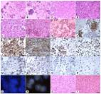
          We present a case of a 22-year-old medical student with a 4-month history of headaches and diplopia. A previous magnetic resonance imaging scan revealed a mass in the pineal region, with heterogeneous contrast enhancement and hydrocephalus. Three months prior, an endoscopic biopsy and third ventriculocisternostomy were performed elsewhere; the diagnosis was neurocytoma Grade I, and radiotherapy was planned. The patient presented at our hospital for a second opinion, and we suggested surgical treatment. A near-total resection was performed in sitting position using a supracerebellar infratentorial microsurgical approach. The tumor was very soft and not well vascularized. Diplopia was initially worsened after the tumor was removed and relieved completely after 2 weeks. An 8-week follow-up examination revealed that the patient was free of symptoms. Histological analysis confirmed it was an RGNT.

          Conclusion:

          Maximal safe resection in pineal region RGNTs is a feasible and recommended treatment option.

          Related collections

          Most cited references12

          • Record: found
          • Abstract: found
          • Article: not found

          A rosette-forming glioneuronal tumor of the fourth ventricle: infratentorial form of dysembryoplastic neuroepithelial tumor?

          Eleven cases of a distinctive tumor of the posterior fossa are described. The patients (age range 12-59 years) presented with headache and/or ataxia. Neuroimaging revealed a relatively discrete, focally enhancing mass(es) primarily involving the aqueduct, fourth ventricle, and cerebellar vermis. Hydrocephalus was present in seven cases, and two lesions were multicentric. In two cases a significant increase in tumor size was documented. Gross total or subtotal resections were achieved in 10 cases. One patient underwent biopsy alone and another received postoperative irradiation. Histologically, two components were identified in all cases. One consisted of neurocytes forming neurocytic and/or perivascular pseudorosettes in a fibrillary, partly microcystic matrix. The second, astrocytic component resembled pilocytic astrocytoma in 10 cases and consisted of fibrillated spindle cells with oval nuclei associated with occasional Rosenthal fibers, granular bodies, glomeruloid capillaries, and microcalcifications. Regionally, this component was more diffuse and patternless, consisting of sheets of round to oval, oligodendrocyte-like cells. Rare ganglion cells were seen in four cases. The rosettes were consistently synaptophysin and MAP-2 immunoreactive, whereas the spindle cells were positive for S-100 protein and glial fibrillary acidic protein. Overall, atypia was minimal; no mitoses were found, and Ki67 labeling indices were low. Ultrastructurally, the neurocytic cells featured processes containing microtubules and occasional dense core granules. Mature synapses were found in one of the four cases studied. Although the histologic features of this unique tumor superficially resemble those of dysembryoplastic neuroepithelial tumor, rosette formation by neuronal cells, the frequent presence of a pilocytic astrocytoma component, and the growing nature of the lesion argue against that diagnosis, as does occasional multifocality.
            Bookmark
            • Record: found
            • Abstract: found
            • Article: not found

            Rosette-forming glioneuronal tumor: report of an unusual case with intraventricular dissemination.

            A rosette-forming glioneuronal tumor (RGNT) was encountered in a 16-year-old Chinese girl. She experienced seizures with loss of consciousness for 1 month prior to diagnosis. A brain MRI revealed multifocal masses occupying all of the ventricular system associated with marked hydrocephalus. A biopsy was performed on the right lateral ventricle using a neuroendoscope and the patient was given postoperative radiotherapy. She was followed for 7 months, and there was no radiological or clinical evidence of tumor progression. Histological examination demonstrated two regions characterized by predominant neurocytic rosettes and scant low grade astrocytoma. No histological anaplasia was present. Immunohistological studies showed that the small round tumor cells forming rosettes were strongly positive for Olig2. A chromosome 1p/19q co-deletion was not detected by FISH analysis. While the pathological features of our case closely resembled those reported in the original description, the diffuse intraventricular growth pattern of the tumor was different from previous examples. Further long-term follow-up studies are required to assess the biological behavior and clinical outcome of the RGNT entity.
              Bookmark
              • Record: found
              • Abstract: found
              • Article: found
              Is Open Access

              Histopathological, molecular, clinical and radiological characterization of rosette-forming glioneuronal tumor in the central nervous system

              Objective A rosette-forming glioneuronal tumor (RGNT) is a rare entity originally described in the fourth ventricle. Recently, RGNTs occurring in extraventricular sites and those with malignant behaviors have been reported. The purpose of this study was to analyze the clinicoradiological and histopathological features, therapeutic strategies, and outcomes of RGNTs. Methods We enrolled 38 patients diagnosed with RGNTs pathologically between August 2009 and June 2016. CT and MRI, including diffusion-weighted imaging and spectroscopy, were performed. The surgical treatment and histopathological and molecular features were assessed. Additionally, we searched the relevant literatures and performed a pooled analysis of individual patient data. The potential risk factors of prognosis were analyzed. Results Our case series included 22 male and 16 female patients, with a mean age of 25.9 years. RGNTs involved the fourth ventricle (26.3%), cerebella (34.2%), supratentorial ventricular system (13.2%), spinal cord (10.5%), temporal lobe (10.5%), thalamus (7.9%), brain stem (7.9%), frontal lobe (5.3%), pineal region (5.3%), suprasellar region (2.6%), and basal ganglia (2.6%). Statistical analyses showed that pediatric age, purely solid appearance of the tumor, and inadequate resection (only partial removal or biopsy) were risk factors associated with progression events. Patients with subtotal resection appeared to do as well as those with gross total resection. Conclusions RGNTs can occur nearly anywhere in the CNS, at both supratentorial and infratentorial sites. Maximal safe surgical resection should be emphasized for treatment; whilst aggressive resection with the goal of complete resection may be unnecessary.
                Bookmark

                Author and article information

                Contributors
                Journal
                Surg Neurol Int
                Surg Neurol Int
                Surgical Neurology International
                Scientific Scholar (USA )
                2229-5097
                2152-7806
                2020
                18 April 2020
                : 11
                : 68
                Affiliations
                [1 ]Department of Neurosurgery, University of Helsinki and Helsinki University Hospital, Helsinki, Finland,
                [2 ]Department of Neurosurgery, Heinrich-Heine University Medical Center, Düsseldorf, Germany.
                Author notes
                [* ] Corresponding author: Sajjad Muhammad, Department of Neurosurgery, University of Helsinki, Topeliuksenkatu 5, Helsinki-FI-000, Helsinki, Finland. ext-sajjad.muhammad@ 123456hus.fi
                Article
                SNI-11-68
                10.25259/SNI_294_2019
                7193215
                32363063
                a76a35cf-5870-4a2c-8a4c-be259ed629e5
                Copyright: © 2020 Surgical Neurology International

                This is an open-access article distributed under the terms of the Creative Commons Attribution-Non Commercial-Share Alike 4.0 License, which allows others to remix, tweak, and build upon the work non-commercially, as long as the author is credited and the new creations are licensed under the identical terms.

                History
                : 08 May 2019
                : 17 March 2020
                Categories
                Case Report

                Surgery
                operative nuances,pineal region,rosette-forming glioneuronal tumor
                Surgery
                operative nuances, pineal region, rosette-forming glioneuronal tumor

                Comments

                Comment on this article