11
views
0
recommends
+1 Recommend
1 collections
    0
    shares

      Drug Design, Development and Therapy (submit here)

      This international, peer-reviewed Open Access journal by Dove Medical Press focuses on the design and development of drugs, as well as the clinical outcomes, patient safety, and programs targeted at the effective and safe use of medicines. Sign up for email alerts here.

      88,007 Monthly downloads/views I 4.319 Impact Factor I 6.6 CiteScore I 1.12 Source Normalized Impact per Paper (SNIP) I 0.784 Scimago Journal & Country Rank (SJR)


      • Record: found
      • Abstract: found
      • Article: found
      Is Open Access

      Annatto-derived tocotrienol stimulates osteogenic activity in preosteoblastic MC3T3-E1 cells: a temporal sequential study

      research-article

      Read this article at

      ScienceOpenPublisherPMC
      Bookmark
          There is no author summary for this article yet. Authors can add summaries to their articles on ScienceOpen to make them more accessible to a non-specialist audience.

          Abstract

          Purpose

          Annatto-derived tocotrienol (AnTT) has been shown to improve bone formation in animal models of osteoporosis. However, detailed studies of the effects of AnTT on preosteoblastic cells were limited. This study was conducted to investigate the osteogenic effect of AnTT on preosteoblast MC3T3-E1 cells in a time-dependent manner.

          Materials and methods

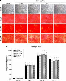
          Murine MC3T3-E1 preosteoblastic cells were cultured in the different concentrations of AnTT (0.001–1 µg/mL) up to 24 days. Expression of osteoblastic differentiation markers was measured by qPCR (osterix [OSX], collagen 1 alpha 1 [COL1α1], alkaline phosphatase [ALP], and osteocalcin [OCN]) and by fluorometric assay for ALP activity. Detection of collagen and mineralized nodules was done via Direct Red staining and Alizarin Red staining, respectively.

          Results

          The results showed that osteoblastic differentiation-related genes, such as OSX, COL1α1, ALP, and OCN, were significantly increased in the AnTT-treated groups compared to the vehicle group in a time-dependent manner ( P<0.05). Type 1 collagen level was increased from day 3 to day 15 in the AnTT-treated groups, while ALP activity was increased from day 9 to day 21 in the AnTT-treated groups ( P<0.05). Enhanced mineralization was observed in the AnTT-treated groups via increasing Alizarin Red staining from day 3 to day 21 ( P<0.05).

          Conclusion

          Our results suggest that AnTT enhances the osteogenic activity by promoting the bone formation-related genes and proteins in a temporal and sequential manner.

          Most cited references32

          • Record: found
          • Abstract: found
          • Article: not found

          In vitro differentiation and calcification in a new clonal osteogenic cell line derived from newborn mouse calvaria

          We investigated the capacity of a clonal osteogenic cell line MC3T3-E1, established from newborn mouse calvaria and selected on the basis of high alkaline phosphatase (ALP) activity in the confluent state, to differentiate into osteoblasts and mineralize in vitro. The cells in the growing state showed a fibroblastic morphology and grew to form multiple layers. On day 21, clusters of cells exhibiting typical osteoblastic morphology were found in osmiophilic nodular regions. Such nodules increased in number and size with incubation time and became easily identifiable with the naked eye by day 40-50. In the central part of well-developed nodules, osteocytes were embedded in heavily mineralized bone matrix. Osteoblasts were arranged at the periphery of the bone spicules and were surrounded by lysosome-rich cells and a fibroblastic cell layer. Numerous matrix vesicles were scattered around the osteoblasts and young osteocytes. Matrix vesicles and plasma membranes of osteoblasts, young osteocytes, and lysosome-rich cells showed strong reaction to cytochemical stainings for ALP activity and calcium ions. Minerals were initially localized in the matrix vesicles and then deposited on well-banded collagen fibrils. Deposited minerals consisted exclusively of calcium and phosphorus, and some of the crystals had matured into hydroxyapatite crystals. These results indicate that MC3T3-E1 cells have the capacity to differentiate into osteoblasts and osteocytes and to form calcified bone tissue in vitro.
            Bookmark
            • Record: found
            • Abstract: found
            • Article: not found

            Progressive development of the rat osteoblast phenotype in vitro: reciprocal relationships in expression of genes associated with osteoblast proliferation and differentiation during formation of the bone extracellular matrix.

            The relationship of cell proliferation to the temporal expression of genes characterizing a developmental sequence associated with bone cell differentiation was examined in primary diploid cultures of fetal calvarial derived osteoblasts by the combined use of autoradiography, histochemistry, biochemistry, and mRNA assays of osteoblast cell growth and phenotypic genes. Modifications in gene expression define a developmental sequence that has 1) three principle periods--proliferation, extracellular matrix maturation, and mineralization--and 2) two restriction points to which the cells can progress but cannot pass without further signals--the first when proliferation is down-regulated and gene expression associated with extracellular matrix maturation is induced, and the second when mineralization occurs. Initially, actively proliferating cells, expressing cell cycle- and cell growth-regulated genes, produce a fibronectin/type I collagen extracellular matrix. A reciprocal and functionally coupled relationship between the decline in proliferative activity and the subsequent induction of genes associated with matrix maturation and mineralization is supported by 1) a temporal sequence of events in which there is an enhanced expression of alkaline phosphatase immediately following the proliferative period, and later, an increased expression of osteocalcin and osteopontin at the onset of mineralization; 2) increased expression of a specific subset of osteoblast phenotype markers, alkaline phosphatase and osteopontin, when proliferation is inhibited by hydroxyurea; and 3) enhanced levels of expression of the osteoblast markers as a function of ascorbic acid-induced collagen deposition, suggesting that the extracellular matrix contributes to both the shutdown of proliferation and the development of the osteoblast phenotype.
              Bookmark
              • Record: found
              • Abstract: found
              • Article: not found

              Distinct proliferative and differentiated stages of murine MC3T3-E1 cells in culture: an in vitro model of osteoblast development.

              We examine clonal murine calvarial MC3T3-E1 cells to determine if they exhibit a developmental sequence similar to osteoblasts in bone tissue, namely, proliferation of undifferentiated osteoblast precursors followed by postmitotic expression of differentiated osteoblast phenotype. During the initial phase of developmental (days 1-9 of culture), MC3T3-E1 cells actively replicate, as evidenced by the high rates of DNA synthesis and progressive increase in cell number, but maintain a fusiform appearance, fail to express alkaline phosphatase, and do not accumulate mineralized extracellular collagenous matrix, consistent with immature osteoblasts. By day 9 the cultures display cuboidal morphology, attain confluence, and undergo growth arrest. Downregulation of replication is associated with expression of osteoblast functions, including production of alkaline phosphatase, processing of procollagens to collagens, and incremental deposition of a collagenous extracellular matrix. Mineralization of extracellular matrix, which begins approximately 16 days after culture, marks the final phase of osteoblast phenotypic development. Expression of alkaline phosphatase and mineralization is time but not density dependent. Type I collagen synthesis and collagen accumulation are uncoupled in the developing osteoblast. Although collagen synthesis and message expression peaks at day 3 in immature cells, extracellular matrix accumulation is minimal. Instead, matrix accumulates maximally after 7 days of culture as collagen biosynthesis is diminishing. Thus, extracellular matrix formation is a function of mature osteoblasts. Ascorbate and beta-glycerol phosphate are both essential for the expression of osteoblast phenotype as assessed by alkaline phosphatase and mineralization of extracellular matrix. Ascorbate does not stimulate type I collagen gene expression in MC3T3-E1 cells, but it is absolutely required for deposition of collagen in the extracellular matrix. Ascorbate also induces alkaline phosphatase activity in mature cells but not in immature cells. beta-glycerol phosphate displays synergistic actions with ascorbate to further stimulate collagen accumulation and alkaline phosphatase activity in postmitotic, differentiated osteoblast-like cells. Mineralization of mature cultures requires the presence of beta-glycerol phosphate. Thus, MC3T3-E1 cells display a time-dependent and sequential expression of osteoblast characteristics analogous to in vivo bone formation. The developmental sequence associated with MC3T3-E1 differentiation should provide a useful model to study the signals that mediate the switch between proliferation and differentiation in bone cells, as well as provide a renewable culture system to examine the molecular mechanism of osteoblast maturation and the formation of bone-like extracellular matrix.
                Bookmark

                Author and article information

                Journal
                Drug Des Devel Ther
                Drug Des Devel Ther
                Drug Design, Development and Therapy
                Drug Design, Development and Therapy
                Dove Medical Press
                1177-8881
                2018
                13 June 2018
                : 12
                : 1715-1726
                Affiliations
                [1 ]Department of Pharmacology, Faculty of Medicine, Universiti Kebangsaan Malaysia Medical Centre, Jalan Yaacob Latif, Bandar Tun Razak, Cheras, Kuala Lumpur, Malaysia
                [2 ]Department of Anatomy, Faculty of Medicine, Universiti Kebangsaan Malaysia Medical Centre, Jalan Yaacob Latif, Bandar Tun Razak, Cheras, Kuala Lumpur, Malaysia
                Author notes
                Correspondence: Soelaiman Ima-Nirwana, Department of Pharmacology, Faculty of Medicine, Universiti Kebangsaan Malaysia Medical Centre, Jalan Yaacob Latif, Bandar Tun Razak, Cheras, 56000 Kuala Lumpur, Malaysia, Tel +60 3 9145 5002, Fax +60 3 9145 6633, Email imasoel@ 123456ppukm.ukm.edu.my
                Article
                dddt-12-1715
                10.2147/DDDT.S168935
                6005313
                a7b3428c-a34a-42e9-83ff-3fabe9f8a1f7
                © 2018 Wan Hasan et al. This work is published and licensed by Dove Medical Press Limited

                The full terms of this license are available at https://www.dovepress.com/terms.php and incorporate the Creative Commons Attribution – Non Commercial (unported, v3.0) License ( http://creativecommons.org/licenses/by-nc/3.0/). By accessing the work you hereby accept the Terms. Non-commercial uses of the work are permitted without any further permission from Dove Medical Press Limited, provided the work is properly attributed.

                History
                Categories
                Original Research

                Pharmacology & Pharmaceutical medicine
                bone,differentiation,osteoporosis,tocotrienol,vitamin e
                Pharmacology & Pharmaceutical medicine
                bone, differentiation, osteoporosis, tocotrienol, vitamin e

                Comments

                Comment on this article