34
views
0
recommends
+1 Recommend
0 collections
    0
    shares
      • Record: found
      • Abstract: found
      • Article: not found

      Quantification, improvement, and harmonization of small lesion detection with state-of-the-art PET

      review-article

      Read this article at

      ScienceOpenPublisherPMC
      Bookmark
          There is no author summary for this article yet. Authors can add summaries to their articles on ScienceOpen to make them more accessible to a non-specialist audience.

          Abstract

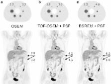
          In recent years, there have been multiple advances in positron emission tomography/computed tomography (PET/CT) that improve cancer imaging. The present generation of PET/CT scanners introduces new hardware, software, and acquisition methods. This review describes these new developments, which include time-of-flight (TOF), point-spread-function (PSF), maximum-a-posteriori (MAP) based reconstruction, smaller voxels, respiratory gating, metal artefact reduction, and administration of quadratic weight-dependent 18F–fluorodeoxyglucose (FDG) activity. Also, hardware developments such as continuous bed motion (CBM), (digital) solid-state photodetectors and combined PET and magnetic resonance (MR) systems are explained. These novel techniques have a significant impact on cancer imaging, as they result in better image quality, improved small lesion detectability, and more accurate quantification of radiopharmaceutical uptake. This influences cancer diagnosis and staging, as well as therapy response monitoring and radiotherapy planning. Finally, the possible impact of these developments on the European Association of Nuclear Medicine (EANM) guidelines and EANM Research Ltd. (EARL) accreditation for FDG-PET/CT tumor imaging is discussed.

          Related collections

          Most cited references74

          • Record: found
          • Abstract: found
          • Article: not found

          FDG PET and PET/CT: EANM procedure guidelines for tumour PET imaging: version 1.0

          The aim of this guideline is to provide a minimum standard for the acquisition and interpretation of PET and PET/CT scans with [18F]-fluorodeoxyglucose (FDG). This guideline will therefore address general information about [18F]-fluorodeoxyglucose (FDG) positron emission tomography-computed tomography (PET/CT) and is provided to help the physician and physicist to assist to carrying out, interpret, and document quantitative FDG PET/CT examinations, but will concentrate on the optimisation of diagnostic quality and quantitative information.
            Bookmark
            • Record: found
            • Abstract: found
            • Article: not found

            Fully 3-D PET reconstruction with system matrix derived from point source measurements.

            The quality of images reconstructed by statistical iterative methods depends on an accurate model of the relationship between image space and projection space through the system matrix The elements of the system matrix for the clinical Hi-Rez scanner were derived by processing the data measured for a point source at different positions in a portion of the field of view. These measured data included axial compression and azimuthal interleaving of adjacent projections. Measured data were corrected for crystal and geometrical efficiency. Then, a whole system matrix was derived by processing the responses in projection space. Such responses included both geometrical and detection physics components of the system matrix. The response was parameterized to correct for point source location and to smooth for projection noise. The model also accounts for axial compression (span) used on the scanner. The forward projector for iterative reconstruction was constructed using the estimated response parameters. This paper extends our previous work to fully three-dimensional. Experimental data were used to compare images reconstructed by the standard iterative reconstruction software and the one modeling the response function. The results showed that the modeling of the response function improves both spatial resolution and noise properties.
              Bookmark
              • Record: found
              • Abstract: found
              • Article: not found

              Phantom and Clinical Evaluation of the Bayesian Penalized Likelihood Reconstruction Algorithm Q.Clear on an LYSO PET/CT System.

              Q.Clear, a Bayesian penalized-likelihood reconstruction algorithm for PET, was recently introduced by GE Healthcare on their PET scanners to improve clinical image quality and quantification. In this work, we determined the optimum penalization factor (beta) for clinical use of Q.Clear and compared Q.Clear with standard PET reconstructions.
                Bookmark

                Author and article information

                Contributors
                +31 6 115 130 27 , eric.visser@radboudumc.nl
                Journal
                Eur J Nucl Med Mol Imaging
                Eur. J. Nucl. Med. Mol. Imaging
                European Journal of Nuclear Medicine and Molecular Imaging
                Springer Berlin Heidelberg (Berlin/Heidelberg )
                1619-7070
                1619-7089
                8 July 2017
                8 July 2017
                2017
                : 44
                : Suppl 1
                : 4-16
                Affiliations
                [1 ]ISNI 0000 0004 0444 9382, GRID grid.10417.33, Department of Radiology and Nuclear Medicine, , Radboud University Medical Centre, ; Nijmegen, The Netherlands
                [2 ]ISNI 0000 0004 0399 8953, GRID grid.6214.1, MIRA Institute for Biomedical Technology and Technical Medicine, , University of Twente, ; Enschede, The Netherlands
                [3 ]ISNI 0000 0001 0547 5927, GRID grid.452600.5, Department of Nuclear Medicine, , Isala Hospital, ; Zwolle, The Netherlands
                [4 ]ISNI 0000 0004 0398 8384, GRID grid.413532.2, Department of Medical Physics, , Catharina Hospital, ; Eindhoven, The Netherlands
                [5 ]Department of Nuclear Medicine & Molecular Imaging, University of Groningen, University Medical Centre Groningen, Groningen, The Netherlands
                [6 ]ISNI 0000 0004 0435 165X, GRID grid.16872.3a, Department of Radiology and Nuclear Medicine, , VU University Medical Center, ; Amsterdam, The Netherlands
                [7 ]ISNI 0000 0001 0547 5927, GRID grid.452600.5, Department of Medical Physics, , Isala, ; Zwolle, The Netherlands
                [8 ]ISNI 0000 0004 1936 9457, GRID grid.8993.b, Department of Surgical Sciences, , Uppsala University, ; Uppsala, Sweden
                [9 ]ISNI 0000 0001 2351 3333, GRID grid.412354.5, Department of Medical Physics, , Uppsala University Hospital, ; Uppsala, Sweden
                Article
                3727
                10.1007/s00259-017-3727-z
                5541089
                28687866
                a7ea4534-7e80-4535-b7af-d07e8d1bf12f
                © The Author(s) 2017

                Open Access This article is distributed under the terms of the Creative Commons Attribution 4.0 International License (http://creativecommons.org/licenses/by/4.0/), which permits unrestricted use, distribution, and reproduction in any medium, provided you give appropriate credit to the original author(s) and the source, provide a link to the Creative Commons license, and indicate if changes were made.

                History
                : 4 May 2017
                : 9 May 2017
                Funding
                Funded by: Radboud University Medical Center
                Categories
                Review Article
                Custom metadata
                © Springer-Verlag GmbH Germany 2017

                Radiology & Imaging
                time-of-flight,point-spread-function,digital pet,pet/mr,lesion detectability,earl
                Radiology & Imaging
                time-of-flight, point-spread-function, digital pet, pet/mr, lesion detectability, earl

                Comments

                Comment on this article