95
views
0
recommends
+1 Recommend
0 collections
    0
    shares
      • Record: found
      • Abstract: found
      • Article: found
      Is Open Access

      Novel Markers to Delineate Murine M1 and M2 Macrophages

      research-article

      Read this article at

      Bookmark
          There is no author summary for this article yet. Authors can add summaries to their articles on ScienceOpen to make them more accessible to a non-specialist audience.

          Abstract

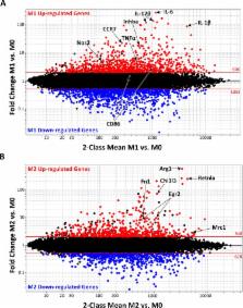
          Classically (M1) and alternatively activated (M2) macrophages exhibit distinct phenotypes and functions. It has been difficult to dissect macrophage phenotypes in vivo, where a spectrum of macrophage phenotypes exists, and also in vitro, where low or non-selective M2 marker protein expression is observed. To provide a foundation for the complexity of in vivo macrophage phenotypes, we performed a comprehensive analysis of the transcriptional signature of murine M0, M1 and M2 macrophages and identified genes common or exclusive to either subset. We validated by real-time PCR an M1-exclusive pattern of expression for CD38, G-protein coupled receptor 18 (Gpr18) and Formyl peptide receptor 2 (Fpr2) whereas Early growth response protein 2 (Egr2) and c-Myc were M2-exclusive. We further confirmed these data by flow cytometry and show that M1 and M2 macrophages can be distinguished by their relative expression of CD38 and Egr2. Egr2 labeled more M2 macrophages (~70%) than the canonical M2 macrophage marker Arginase-1, which labels 24% of M2 macrophages. Conversely, CD38 labeled most (71%) in vitro M1 macrophages. In vivo, a similar CD38 + population greatly increased after LPS exposure. Overall, this work defines exclusive and common M1 and M2 signatures and provides novel and improved tools to distinguish M1 and M2 murine macrophages.

          Related collections

          Most cited references45

          • Record: found
          • Abstract: found
          • Article: not found

          Transcriptional profiling of the human monocyte-to-macrophage differentiation and polarization: new molecules and patterns of gene expression.

          Comprehensive analysis of the gene expression profiles associated with human monocyte-to-macrophage differentiation and polarization toward M1 or M2 phenotypes led to the following main results: 1) M-CSF-driven monocyte-to-macrophage differentiation is associated with activation of cell cycle genes, substantiating the underestimated proliferation potential of monocytes. 2) M-CSF leads to expression of a substantial part of the M2 transcriptome, suggesting that under homeostatic conditions a default shift toward M2 occurs. 3) Modulation of genes involved in metabolic activities is a prominent feature of macrophage differentiation and polarization. 4) Lipid metabolism is a main category of modulated transcripts, with expected up-regulation of cyclo-oxygenase 2 in M1 cells and unexpected cyclo-oxygenase 1 up-regulation in M2 cells. 5) Each step is characterized by a different repertoire of G protein-coupled receptors, with five nucleotide receptors as novel M2-associated genes. 6) The chemokinome of polarized macrophages is profoundly diverse and new differentially expressed chemokines are reported. Thus, transcriptome profiling reveals novel molecules and signatures associated with human monocyte-to-macrophage differentiation and polarized activation which may represent candidate targets in pathophysiology.
            Bookmark
            • Record: found
            • Abstract: found
            • Article: not found

            Cell type-specific involvement of RIG-I in antiviral response.

            Toll-like receptors (TLRs) play an important role in antiviral response by recognizing viral components. Recently, a RNA helicase, RIG-I, was also suggested to recognize viral double-stranded RNA. However, how these molecules contribute to viral recognition in vivo is poorly understood. We show by gene targeting that RIG-I is essential for induction of type I interferons (IFNs) after infection with RNA viruses in fibroblasts and conventional dendritic cells (DCs). RIG-I induces type I IFNs by activating IRF3 via IkappaB kinase-related kinases. In contrast, plasmacytoid DCs, which produce large amounts of IFN-alpha, use the TLR system rather than RIG-I for viral detection. Taken together, RIG-I and the TLR system exert antiviral responses in a cell type-specific manner.
              Bookmark
              • Record: found
              • Abstract: found
              • Article: not found

              Macrophage polarization in bacterial infections.

              Converging studies have shown that M1 and M2 macrophages are functionally polarized in response to microorganisms and host mediators. Gene expression profiling of macrophages reveals that various Gram-negative and Gram-positive bacteria induce the transcriptional activity of a "common host response," which includes genes belonging to the M1 program. However, excessive or prolonged M1 polarization can lead to tissue injury and contribute to pathogenesis. The so-called M2 macrophages play a critical role in the resolution of inflammation by producing anti-inflammatory mediators. These M2 cells cover a continuum of cells with different phenotypic and functional properties. In addition, some bacterial pathogens induce specific M2 programs in macrophages. In this review, we discuss the relevance of macrophage polarization in three domains of infectious diseases: resistance to infection, infectious pathogenesis, and chronic evolution of infectious diseases.
                Bookmark

                Author and article information

                Contributors
                Role: Editor
                Journal
                PLoS One
                PLoS ONE
                plos
                plosone
                PLoS ONE
                Public Library of Science (San Francisco, CA USA )
                1932-6203
                23 December 2015
                2015
                : 10
                : 12
                : e0145342
                Affiliations
                [1 ]School of Health and Rehabilitation Sciences, Medical Laboratory Science Division, The Ohio State University, Columbus, Ohio, United States of America
                [2 ]Biomedical Sciences Graduate Program, The Ohio State University, Columbus, Ohio, United States of America
                [3 ]Center for Microbial Pathogenesis, The Research Institute at Nationwide Children’s Hospital, Columbus, Ohio, United States of America
                [4 ]Department of Neuroscience, Wexner Medical Center at The Ohio State University, Columbus, Ohio, United States of America
                University of Michigan Health System, UNITED STATES
                Author notes

                Competing Interests: The authors have declared that no competing interests exist.

                Conceived and designed the experiments: KJ SA LW JRR PP SPS MG. Performed the experiments: KJ SA LW JRR MG. Analyzed the data: KJ SA LW JRR MG. Contributed reagents/materials/analysis tools: PP SPS MG. Wrote the paper: KJ SA LW JRR PP SPS MG.

                Article
                PONE-D-15-42425
                10.1371/journal.pone.0145342
                4689374
                26699615
                a8e4db0d-0674-4eac-b1a2-b08f5181cde1
                © 2015 Jablonski et al

                This is an open access article distributed under the terms of the Creative Commons Attribution License, which permits unrestricted use, distribution, and reproduction in any medium, provided the original author and source are credited

                History
                : 25 September 2015
                : 2 December 2015
                Page count
                Figures: 7, Tables: 5, Pages: 25
                Funding
                This work was supported by funds from the International Foundation for Research on Paraplegia (P 129 to PP and MG), the National Institutes of Health (NIH R21 R21NS081413 to PP and MG and NIAID R01-AI092117 to SP-S), and the Ray W. Poppleton Endowment (PP). KJ was supported by the Ohio State University Mayer’s Undergraduate Summer Research Fellowship and LW by the Systems and Integrative Biology Fellowship (NIH T32). The funders had no role in study design, data collection and analysis, decision to publish, or preparation of the manuscript.
                Categories
                Research Article
                Custom metadata
                Array data are deposited at the Gene Expression Omnibus (GEO) NCBI database with accession number (GSE69607).

                Uncategorized
                Uncategorized

                Comments

                Comment on this article