6
views
0
recommends
+1 Recommend
0 collections
    0
    shares
      • Record: found
      • Abstract: found
      • Article: found
      Is Open Access

      Age-related abnormalities of thalamic shape and dynamic functional connectivity after three hours of sleep restriction

      research-article

      Read this article at

      Bookmark
          There is no author summary for this article yet. Authors can add summaries to their articles on ScienceOpen to make them more accessible to a non-specialist audience.

          Abstract

          Background

          Previous neuroimaging studies have detected abnormal activation and intrinsic functional connectivity of the thalamus after total sleep deprivation. However, very few studies have investigated age-related changes in the dynamic functional connectivity of the thalamus and the abnormalities in the thalamic shape following partial sleep deprivation.

          Methods

          Fifty-five participants consisting of 23 old adults (mean age: 68.8 years) and 32 young adults (mean age: 23.5 years) were included in current study. A vertex-based shape analysis and a dynamic functional connectivity analysis were used to evaluate the age-dependent structural and functional abnormalities after three hours of sleep restriction.

          Results

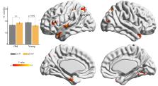
          Shape analysis revealed the significant main effect of deprivation with local atrophy in the left thalamus. In addition, we observed a significant age deprivation interaction effect with reduced variability of functional connectivity between the left thalamus and the left superior parietal cortex following sleep restriction. This reduction was found only in young adults. Moreover, a significantly negative linear correlation was observed between the insomnia severity index and the changes of variability (post-deprivation minus pre-deprivation) in the functional connectivity of the left thalamus with the left superior parietal cortex.

          Conclusions

          The results indicated that three hours of sleep restriction could affect both the thalamic structure and its functional dynamics. They also highlighted the role of age in studies of sleep deprivation.

          Related collections

          Most cited references44

          • Record: found
          • Abstract: found
          • Article: not found

          Automated anatomical labeling of activations in SPM using a macroscopic anatomical parcellation of the MNI MRI single-subject brain.

          An anatomical parcellation of the spatially normalized single-subject high-resolution T1 volume provided by the Montreal Neurological Institute (MNI) (D. L. Collins et al., 1998, Trans. Med. Imag. 17, 463-468) was performed. The MNI single-subject main sulci were first delineated and further used as landmarks for the 3D definition of 45 anatomical volumes of interest (AVOI) in each hemisphere. This procedure was performed using a dedicated software which allowed a 3D following of the sulci course on the edited brain. Regions of interest were then drawn manually with the same software every 2 mm on the axial slices of the high-resolution MNI single subject. The 90 AVOI were reconstructed and assigned a label. Using this parcellation method, three procedures to perform the automated anatomical labeling of functional studies are proposed: (1) labeling of an extremum defined by a set of coordinates, (2) percentage of voxels belonging to each of the AVOI intersected by a sphere centered by a set of coordinates, and (3) percentage of voxels belonging to each of the AVOI intersected by an activated cluster. An interface with the Statistical Parametric Mapping package (SPM, J. Ashburner and K. J. Friston, 1999, Hum. Brain Mapp. 7, 254-266) is provided as a freeware to researchers of the neuroimaging community. We believe that this tool is an improvement for the macroscopical labeling of activated area compared to labeling assessed using the Talairach atlas brain in which deformations are well known. However, this tool does not alleviate the need for more sophisticated labeling strategies based on anatomical or cytoarchitectonic probabilistic maps.
            Bookmark
            • Record: found
            • Abstract: found
            • Article: not found

            DPABI: Data Processing & Analysis for (Resting-State) Brain Imaging.

            Brain imaging efforts are being increasingly devoted to decode the functioning of the human brain. Among neuroimaging techniques, resting-state fMRI (R-fMRI) is currently expanding exponentially. Beyond the general neuroimaging analysis packages (e.g., SPM, AFNI and FSL), REST and DPARSF were developed to meet the increasing need of user-friendly toolboxes for R-fMRI data processing. To address recently identified methodological challenges of R-fMRI, we introduce the newly developed toolbox, DPABI, which was evolved from REST and DPARSF. DPABI incorporates recent research advances on head motion control and measurement standardization, thus allowing users to evaluate results using stringent control strategies. DPABI also emphasizes test-retest reliability and quality control of data processing. Furthermore, DPABI provides a user-friendly pipeline analysis toolkit for rat/monkey R-fMRI data analysis to reflect the rapid advances in animal imaging. In addition, DPABI includes preprocessing modules for task-based fMRI, voxel-based morphometry analysis, statistical analysis and results viewing. DPABI is designed to make data analysis require fewer manual operations, be less time-consuming, have a lower skill requirement, a smaller risk of inadvertent mistakes, and be more comparable across studies. We anticipate this open-source toolbox will assist novices and expert users alike and continue to support advancing R-fMRI methodology and its application to clinical translational studies.
              Bookmark
              • Record: found
              • Abstract: found
              • Article: not found

              Tracking whole-brain connectivity dynamics in the resting state.

              Spontaneous fluctuations are a hallmark of recordings of neural signals, emergent over time scales spanning milliseconds and tens of minutes. However, investigations of intrinsic brain organization based on resting-state functional magnetic resonance imaging have largely not taken into account the presence and potential of temporal variability, as most current approaches to examine functional connectivity (FC) implicitly assume that relationships are constant throughout the length of the recording. In this work, we describe an approach to assess whole-brain FC dynamics based on spatial independent component analysis, sliding time window correlation, and k-means clustering of windowed correlation matrices. The method is applied to resting-state data from a large sample (n = 405) of young adults. Our analysis of FC variability highlights particularly flexible connections between regions in lateral parietal and cingulate cortex, and argues against a labeling scheme where such regions are treated as separate and antagonistic entities. Additionally, clustering analysis reveals unanticipated FC states that in part diverge strongly from stationary connectivity patterns and challenge current descriptions of interactions between large-scale networks. Temporal trends in the occurrence of different FC states motivate theories regarding their functional roles and relationships with vigilance/arousal. Overall, we suggest that the study of time-varying aspects of FC can unveil flexibility in the functional coordination between different neural systems, and that the exploitation of these dynamics in further investigations may improve our understanding of behavioral shifts and adaptive processes.
                Bookmark

                Author and article information

                Contributors
                Journal
                PeerJ
                PeerJ
                peerj
                peerj
                PeerJ
                PeerJ Inc. (San Diego, USA )
                2167-8359
                26 January 2021
                2021
                : 9
                : e10751
                Affiliations
                [1 ]Sleep and NeuroImaging Center, Faculty of Psychology, University of the Southwest , Chongqing, China
                [2 ]Key Laboratory of Cognition and Personality (Southwest University), Ministry of Education , Chongqing, China
                Article
                10751
                10.7717/peerj.10751
                7845526
                aaf424e9-d3c6-4b41-b219-c180fa52ccfc
                ©2021 Long et al.

                This is an open access article distributed under the terms of the Creative Commons Attribution License, which permits unrestricted use, distribution, reproduction and adaptation in any medium and for any purpose provided that it is properly attributed. For attribution, the original author(s), title, publication source (PeerJ) and either DOI or URL of the article must be cited.

                History
                : 9 September 2020
                : 19 December 2020
                Funding
                Funded by: National Natural Science Foundation of China
                Award ID: 81901725
                Funded by: Fundamental Research Funds for the Central Universities
                Award ID: SWU118004
                This work was supported by the National Natural Science Foundation of China (81901725), and the Fundamental Research Funds for the Central Universities (No. SWU118004). The funders had no role in study design, data collection and analysis, decision to publish, or preparation of the manuscript.
                Categories
                Neuroscience
                Anatomy and Physiology
                Neurology
                Psychiatry and Psychology

                sleep loss,aging,thalamus,subcortical shape,functional connectivity variability

                Comments

                Comment on this article