3
views
0
recommends
+1 Recommend
0 collections
    0
    shares
      • Record: found
      • Abstract: found
      • Article: found
      Is Open Access

      Exogenous bone marrow derived-putative endothelial progenitor cells attenuate ischemia reperfusion-induced vascular injury and renal fibrosis in mice dependent on pericytes

      research-article

      Read this article at

      Bookmark
          There is no author summary for this article yet. Authors can add summaries to their articles on ScienceOpen to make them more accessible to a non-specialist audience.

          Abstract

          Rationale: Capillaries are composed of endothelial cells and the surrounding mural cells, pericytes. Microvascular repair after injury involves not only the proliferation of endothelial cells but also pericyte-based vessel stabilization. Exogenous bone marrow derived-putative endothelial progenitor cells (b-pEPCs) have the potential for vascular repair; however, their effect on vascular structure stabilization and pericyte-related pathobiological outcomes in the injured kidney has not been fully examined.

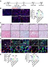
          Methods: We applied ischemia-reperfusion (IR) to induce renal vascular injury and renal fibrosis in mice. Platelet-derived growth factor receptor β (PDGFR-β)-DTR-positive mice were generated to deplete pericytes, and exogenous b-pEPCs and the PDGFR-β ligand, PDGF chain B (PDGF-BB), were employed to explore the relationship among b-pEPCs, pericytes, vascular repair, and early renal fibrosis.

          Results: Administration of b-pEPCs reduced IR-induced pericyte-endothelial detachment, pericyte proliferation, and myofibroblast transition via a paracrine mode, which preserved not only vascular stabilization but also ameliorated IR-initiated renal fibrosis. PDGF-BB upregulated the expression of PDGFR-β, exacerbated vascular abnormality, and pericyte-myofibroblast transition, which were ameliorated by b-pEPCs administration. The exogenous b-pEPCs and their culture medium (CM) induced vascular injury protection, and renal fibrosis was blocked by selective deletion of pericytes.

          Conclusion: Exogenous b-pEPCs directly protect against IR-induced vascular injury and prevent renal fibrosis by inhibiting the activation of PDGFR-β-positive pericytes.

          Related collections

          Most cited references38

          • Record: found
          • Abstract: found
          • Article: not found

          Pericytes: developmental, physiological, and pathological perspectives, problems, and promises.

          Pericytes, the mural cells of blood microvessels, have recently come into focus as regulators of vascular morphogenesis and function during development, cardiovascular homeostasis, and disease. Pericytes are implicated in the development of diabetic retinopathy and tissue fibrosis, and they are potential stromal targets for cancer therapy. Some pericytes are probably mesenchymal stem or progenitor cells, which give rise to adipocytes, cartilage, bone, and muscle. However, there is still confusion about the identity, ontogeny, and progeny of pericytes. Here, we review the history of these investigations, indicate emerging concepts, and point out problems and promise in the field of pericyte biology. Copyright © 2011 Elsevier Inc. All rights reserved.
            Bookmark
            • Record: found
            • Abstract: found
            • Article: not found

            Epithelial cell cycle arrest in G2/M mediates kidney fibrosis after injury.

            Fibrosis is responsible for chronic progressive kidney failure, which is present in a large number of adults in the developed world. It is increasingly appreciated that acute kidney injury (AKI), resulting in aberrant incomplete repair, is a major contributor to chronic fibrotic kidney disease. The mechanism that triggers the fibrogenic response after injury is not well understood. In ischemic, toxic and obstructive models of AKI, we demonstrate a causal association between epithelial cell cycle G2/M arrest and a fibrotic outcome. G2/M-arrested proximal tubular cells activate c-jun NH(2)-terminal kinase (JNK) signaling, which acts to upregulate profibrotic cytokine production. Treatment with a JNK inhibitor, or bypassing the G2/M arrest by administration of a p53 inhibitor or the removal of the contralateral kidney, rescues fibrosis in the unilateral ischemic injured kidney. Hence, epithelial cell cycle arrest at G2/M and its subsequent downstream signaling are hitherto unrecognized therapeutic targets for the prevention of fibrosis and interruption of the accelerated progression of kidney disease.
              Bookmark
              • Record: found
              • Abstract: found
              • Article: not found

              Chronic hypoxia as a mechanism of progression of chronic kidney diseases: from hypothesis to novel therapeutics.

              In chronic kidney disease, functional impairment correlates with tubulointerstitial fibrosis characterised by inflammation, accumulation of extracellular matrix, tubular atrophy and rarefaction of peritubular capillaries. Loss of the microvasculature implies a hypoxic milieu and suggested an important role for hypoxia when the "chronic hypoxia hypothesis" was proposed a decade ago as an explanation for the progressive nature of fibrosis. Recent data in man provide evidence of decreased renal oxygenation in chronic kidney disease while more direct support for a causal role comes from data in rodent models showing that the decline in renal oxygenation precedes matrix accumulation, suggesting hypoxia may both initiate and promote the fibrotic response. Indeed, in vitro studies show that hypoxia can induce pro-fibrotic changes in tubulointerstitial cells. Additional postulated roles for hypoxia in chronic kidney disease are the sustaining of the inflammatory response, the recruitment, retention and differentiation towards a pro-fibrotic phenotype of circulating progenitor cells and the alteration of the function of intrinsic stem cell populations. Given that accumulating data suggests that chronic hypoxia is a final common pathway to end-stage renal disease, therapeutic strategies that target hypoxia may be of benefit in retarding progression. Normalisation of microvascular tone, administration of pro-angiogenic factors to restore microvasculature integrity, activation of hypoxia-inducible transcription factors and hypoxia-mediated targeting and mobilisation of progenitor cells are all potential targets for future therapy. The limited success of existing strategies in retarding chronic kidney disease mandates that these new avenues of treatment be explored.
                Bookmark

                Author and article information

                Journal
                Theranostics
                Theranostics
                thno
                Theranostics
                Ivyspring International Publisher (Sydney )
                1838-7640
                2020
                25 October 2020
                : 10
                : 26
                : 12144-12157
                Affiliations
                [1 ]Nephrology, Tongji Hospital, Tongji Medical College, Huazhong University of Science and Technology, 1095 Jiefang Ave, Wuhan, Hubei, 430030, China.
                [2 ]Geriatrics, Tongji Hospital, Tongji Medical College, Huazhong University of Science and Technology, 1095 Jiefang Ave, Wuhan, Hubei, 430030, China.
                [3 ]Nutrition, Tongji Hospital, Tongji Medical College, Huazhong University of Science and Technology, 1095 Jiefang Ave, Wuhan, Hubei, 430030, China.
                Author notes
                ✉ Corresponding author: Rui Zeng 1, E-mail: zengr126@ 123456126.com ; Ying Yao 1,3, E-mail: yaoyingkk@ 123456126.com ; Juan Yang 1, E-mail: amy19861203@ 123456126.com .

                *Meng Wang and Huzi Xu contributed equally to this work.

                Competing Interests: The authors have declared that no competing interest exists.

                Article
                thnov10p12144
                10.7150/thno.48562
                7667688
                33204334
                ab052e8d-7e97-4919-9dec-345edb7a53bb
                © The author(s)

                This is an open access article distributed under the terms of the Creative Commons Attribution License ( https://creativecommons.org/licenses/by/4.0/). See http://ivyspring.com/terms for full terms and conditions.

                History
                : 23 May 2020
                : 14 October 2020
                Categories
                Research Paper

                Molecular medicine
                pdgf-bb/pdgfr-β,pericyte,pepcs,vascular injury,renal fibrosis.
                Molecular medicine
                pdgf-bb/pdgfr-β, pericyte, pepcs, vascular injury, renal fibrosis.

                Comments

                Comment on this article