10
views
0
recommends
+1 Recommend
0 collections
    0
    shares
      • Record: found
      • Abstract: found
      • Article: found
      Is Open Access

      Denaturation of proteins by surfactants studied by the Taylor dispersion analysis

      research-article

      Read this article at

      Bookmark
          There is no author summary for this article yet. Authors can add summaries to their articles on ScienceOpen to make them more accessible to a non-specialist audience.

          Abstract

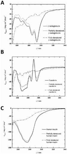
          We showed that the Taylor Dispersion Analysis (TDA) is a fast and easy to use method for the study of denaturation proteins. We applied TDA to study denaturation of β-lactoglobulin, transferrin, and human insulin by anionic surfactant sodium dodecyl sulfate (SDS). A series of measurements at constant protein concentration (for transferrin was 1.9 x 10 −5 M, for β- lactoglobulin was 7.6 x 10 −5 M, and for insulin was 1.2 x 10 −4 M) and varying SDS concentrations were carried out in the phosphate-buffered saline (PBS). The structural changes were analyzed based on the diffusion coefficients of the complexes formed at various surfactant concentrations. The concentration of surfactant was varied in the range from 1.2 x 10 −4 M to 8.7 x 10 −2 M. We determined the minimum concentration of the surfactant necessary to change the native conformation of the proteins. The minimal concentration of SDS for β-lactoglobulin and transferrin was 4.3 x 10 −4 M and for insulin 2.3 x 10 −4 M. To evaluate the TDA as a novel method for studying denaturation of proteins we also applied other methods i.e. electronic circular dichroism (ECD) and dynamic light scattering (DLS) to study the same phenomenon. The results obtained using these methods were in agreement with the results from TDA.

          Related collections

          Most cited references36

          • Record: found
          • Abstract: found
          • Article: not found

          Protein unfolding in detergents: effect of micelle structure, ionic strength, pH, and temperature.

          The 101-residue monomeric protein S6 unfolds in the anionic detergent sodium dodecyl sulfate (SDS) above the critical micelle concentration, with unfolding rates varying according to two different modes. Our group has proposed that spherical micelles lead to saturation kinetics in unfolding (mode 1), while cylindrical micelles prevalent at higher SDS concentrations induce a power-law dependent increase in the unfolding rate (mode 2). Here I investigate in more detail how micellar properties affect protein unfolding. High NaCl concentrations, which induce cylindrical micelles, favor mode 2. This is consistent with our model, though other effects such as electrostatic screening cannot be discounted. Furthermore, unfolding does not occur in mode 2 in the cationic detergent LTAB, which is unable to form cylindrical micelles. A strong retardation of unfolding occurs at higher LTAB concentrations, possibly due to the formation of dead-end protein-detergent complexes. A similar, albeit much weaker, effect is seen in SDS in the absence of salt. Chymotrypsin inhibitor 2 exhibits the same modes of unfolding in SDS as S6, indicating that this type of protein unfolding is not specific for S6. The unfolding process in mode 1 has an activation barrier similar in magnitude to that in water, while the activation barrier in mode 2 is strongly concentration-dependent. The strong pH-dependence of unfolding in SDS and LTAB suggests that the rate of unfolding in anionic detergent is modulated by repulsion between detergent headgroups and anionic side chains, while cationic side chains modulate unfolding rates in cationic detergents.
            Bookmark
            • Record: found
            • Abstract: found
            • Article: not found

            Simple detection of protein soft structure changes.

            We present a rapid method for protein tertiary structure analysis which avoids the need for techniques such as circular dichroism and differential scanning calorimetry. Small changes to a protein's noncovalent "soft" structure are detected by exploiting differences in thermal stability and fluorescent reporter binding. It can detect subtle stability differences using micrograms of protein in 2 microL volumes within minutes.
              Bookmark
              • Record: found
              • Abstract: found
              • Article: not found

              Circular dichroism spectroscopy as a tool for monitoring aggregation in monoclonal antibody therapeutics.

              Aggregation continues to be a critical quality attribute for a monoclonal antibody therapeutic product due to its perceived significant impact on immunogenicity. This paper aims to establish the versatility of circular dichorism (CD) spectroscopy toward understanding aggregation of monoclonal antibody (mAb) therapeutics. The first application involves the use of far-UV CD as a complementary analytical technique to size exclusion chromatography (SEC) for understanding protein aggregation. The second application uses thermal scanning CD as a high throughput screening tool for examining stability of a mAb therapeutic in various formulation and downstream buffers. For establishing far-UV CD as an orthogonal technique, a mAb was incubated in different downstream processing buffers and another mAb in formulation buffers, and they were analyzed by SEC and far-UV CD for aggregate content and conformational stability, respectively. To examine thermal scanning as a high throughput screening tool, ellipticity as a function of the temperature was measured at 218 nm from 20 to 90 °C. Far-UV CD was found to display high sensitivity toward early detection of conformational changes in mAb. CD measurements were also able to elucidate the different aggregation mechanisms. Furthermore, thermal stability scan allowed us to estimate T(onset) which has been found to correlate with aggregation induced by salt, low pH, and buffer species. T(onset) temperature from thermal scanning at 218 nm using CD was correlated successfully to aggregate content measured by SEC. Results from both the studies demonstrate the usefulness of CD for assessing stability of therapeutic proteins during process development, formulation development, and product characterization.
                Bookmark

                Author and article information

                Contributors
                Role: Editor
                Journal
                PLoS One
                PLoS ONE
                plos
                plosone
                PLoS ONE
                Public Library of Science (San Francisco, CA USA )
                1932-6203
                20 April 2017
                2017
                : 12
                : 4
                : e0175838
                Affiliations
                [1 ]Institute of Physical Chemistry, Polish Academy of Sciences, Warsaw, Poland
                [2 ]Institute of Organic Chemistry, Polish Academy of Sciences, Warsaw, Poland
                Russian Academy of Medical Sciences, RUSSIAN FEDERATION
                Author notes

                Competing Interests: The authors have declared that no competing interests exist.

                • Conceptualization: AJ AZ RH.

                • Data curation: AJ AZ.

                • Formal analysis: AJ AZ MG AW.

                • Funding acquisition: RH.

                • Methodology: AJ AZ.

                • Project administration: AJ AZ RH.

                • Resources: AJ AZ MG AW.

                • Software: AJ AZ MG AW.

                • Supervision: RH JF.

                • Validation: AJ AZ MG AW.

                • Visualization: AJ AZ.

                • Writing – original draft: AJ AZ MG AW.

                • Writing – review & editing: AJ.

                Author information
                http://orcid.org/0000-0002-3211-4286
                Article
                PONE-D-17-01178
                10.1371/journal.pone.0175838
                5398553
                28426809
                abb9d5ff-4de7-4ab3-8400-1ab2b495d3b0
                © 2017 Jelińska et al

                This is an open access article distributed under the terms of the Creative Commons Attribution License, which permits unrestricted use, distribution, and reproduction in any medium, provided the original author and source are credited.

                History
                : 10 January 2017
                : 1 April 2017
                Page count
                Figures: 3, Tables: 2, Pages: 11
                Funding
                Funded by: funder-id http://dx.doi.org/10.13039/501100004281, Narodowe Centrum Nauki;
                Award ID: UMO-2012/07/B/ST4/01400
                Award Recipient :
                This research was supported by the National Science Centre Grant Opus 4 (UMO-2012/07/B/ST4/01400).
                Categories
                Research Article
                Physical Sciences
                Materials Science
                Materials by Attribute
                Surfactants
                Biology and life sciences
                Molecular biology
                Molecular biology techniques
                Denaturation
                Protein denaturation
                SDS denaturation techniques
                Research and analysis methods
                Molecular biology techniques
                Denaturation
                Protein denaturation
                SDS denaturation techniques
                Physical Sciences
                Chemistry
                Chemical Physics
                Mass Diffusivity
                Physical Sciences
                Physics
                Chemical Physics
                Mass Diffusivity
                Medicine and Health Sciences
                Endocrinology
                Diabetic Endocrinology
                Insulin
                Biology and Life Sciences
                Biochemistry
                Hormones
                Insulin
                Research and Analysis Methods
                Spectrum Analysis Techniques
                Circular Dichroism Spectroscopy
                Research and analysis methods
                Spectrum analysis techniques
                NMR spectroscopy
                Correlation Spectroscopy
                Photon Correlation Spectroscopy
                Biology and Life Sciences
                Molecular Biology
                Macromolecular Structure Analysis
                Protein Structure
                Biology and Life Sciences
                Biochemistry
                Proteins
                Protein Structure
                Biology and Life Sciences
                Biochemistry
                Proteins
                Structural Proteins
                Custom metadata
                All relevant data are within the paper and its Supporting Information files.

                Uncategorized
                Uncategorized

                Comments

                Comment on this article